| disease | Postpartum Metrorrhagia |
| alias | Postpartum Hemorrhage |
Excessive vaginal bleeding exceeding 500ml within 24 hours after fetal delivery is called postpartum metrorrhagia (postpartum hemorrhage). Postpartum metrorrhagia includes three periods: from fetal delivery to placental delivery, from placental delivery to 2 hours postpartum, and from 2 hours to 24 hours postpartum, with most cases occurring in the first two periods. Postpartum metrorrhagia is one of the leading causes of maternal mortality and currently ranks first in China. Once postpartum metrorrhagia occurs, the prognosis is severe. In cases of prolonged and severe shock, even if the mother is rescued, serious sequelae such as hypopituitarism (Sheehan syndrome) may still occur. Therefore, special attention must be paid to prevention and treatment.
bubble_chart Etiology
It can be divided into four categories: uterine contraction lack of strength, birth canal lacerations, placental factors, and coagulation dysfunction.
1. Uterine contraction lack of strength: After the delivery of the fetus, the placenta separates from the uterine wall and is expelled, leading to the opening of maternal uterine wall blood sinuses and subsequent bleeding. Under normal circumstances, the postpartum uterine cavity volume decreases, and the contraction of muscle fibers strengthens, compressing the blood vessels within the intertwined muscle fibers of the uterine wall to stop bleeding. Simultaneously, the blood sinuses close, and bleeding ceases. Additionally, due to the hypercoagulable state of the maternal blood, platelets aggregate in large numbers on the endothelial collagen fibers of injured blood vessels after placental separation, forming thrombi. Fibrin deposits on the platelet thrombi, creating larger blood clots that effectively block uterine vessels, preventing further bleeding even when muscle fibers relax after contraction. If uterine contraction lack of strength occurs after the delivery of the fetus, the uterus cannot contract and retract normally. If the placenta has not separated and the blood sinuses have not opened, bleeding may not occur. However, if the placenta is partially separated or expelled, uterine contraction lack of strength fails to effectively close the blood sinuses at the placental attachment site, leading to excessive bleeding, which is the main cause of postpartum metrorrhagia.
Uterine contraction lack of strength can result from excessive maternal stress, overuse of sedatives or anesthetics during childbirth; abnormal cephalic presentation or other obstructive difficult deliveries leading to prolonged labor and maternal exhaustion; poor development of uterine muscle fibers in the mother; overdistension of the uterus, such as in cases of twins, macrosomia, or polyhydramnios, causing excessive stretching of uterine muscle fibers; maternal anemia, pregnancy-induced hypertension, or pregnancy complicated by uterine fibroids, all of which can affect uterine contractions.2. Soft birth canal lacerations: Another significant cause of postpartum metrorrhagia. Excessive uterine contraction force, rapid labor progression, or a large fetus can often lead to cervical and/or vaginal lacerations before the fetus is delivered. Improper perineal protection or incorrect delivery assistance techniques can also cause perineal and vaginal lacerations. An episiotomy that is too small may result in severe perineal lacerations during delivery, while premature lateral episiotomy can lead to excessive bleeding from the incision.
Severe perineal and vaginal lacerations can extend upward to the fornix, paravaginal space, or even deep into the pelvic wall. Severe lacerations in the deep vagina near the fornix can lead to hematomas that spread upward into the broad ligament.
During childbirth, minor cervical lacerations are almost inevitable, typically superficial and without significant bleeding, and thus not diagnosed as cervical lacerations. Cervical lacerations with more bleeding occur when the fetus passes too quickly through an incompletely dilated cervix. In severe cases, these lacerations can extend downward to the vaginal fornix or upward to the lower uterine segment, causing massive bleeding.
3. Placental factors: Postpartum metrorrhagia caused by placental factors includes incomplete placental separation, retained placenta after separation, placental incarceration, placental adhesion, placental implantation, and retained placental tissue and/or membranes.Partial placental separation or retained placenta after separation can result from uterine contraction lack of strength. Placental incarceration occasionally occurs after the use of oxytocin or ergot alkaloids, causing a spastic contraction near the internal cervical os, forming a constriction ring that traps the already separated placenta in the uterine cavity, hindering uterine contraction and leading to bleeding. This constriction ring can also occur during rough uterine massage. Overdistension of the bladder can also impede placental expulsion, increasing bleeding.
When the placenta adheres entirely or partially to the uterine wall and cannot separate spontaneously, it is called placental adhesion. Partial adhesion is prone to cause bleeding. Multiple artificial late abortions can easily damage the uterine lining and lead to endometritis. Endometritis can also result from other infections and may cause placental adhesion.
Placental implantation refers to the penetration of placental villi into the uterine muscle layer due to poor development of the uterine decidua or other reasons, which is clinically rare. It can be further classified as complete or partial based on the extent of implantation.
Retained placenta is more common and can result from premature traction of the umbilical cord or excessive manual compression of the uterus. Retained placenta may involve partial placental lobes or accessory placenta adhering to the uterine wall, affecting uterine contractions and causing bleeding. Retained placenta may also include partial retention of membranes.
4. Coagulation dysfunction is a relatively rare cause of postpartum metrorrhagia. Conditions such as hematologic disorders (thrombocytopenia, leukemia, deficiency of coagulation factors VII and VIII, aplastic anemia, etc.) usually exist before pregnancy and are contraindications for pregnancy. Severe hepatitis, prolonged retention of intrauterine dead fetus, placental abruption, grade III pregnancy-induced hypertension, and amniotic fluid embolism can all affect coagulation or lead to disseminated intravascular coagulation, resulting in coagulation disorders, postpartum bleeding with unclotted blood, and difficulty in achieving hemostasis.bubble_chart Clinical Manifestations
The main clinical manifestations of postpartum metrorrhagia are excessive vaginal bleeding, with blood loss exceeding 500ml within 24 hours after childbirth, leading to secondary hemorrhagic shock and susceptibility to infection. The clinical presentation varies depending on the {|###|}disease cause{|###|}.
1. Uterine atony Uterine atony often occurs during childbirth and persists after the delivery of the fetus, though exceptions exist. The bleeding is characterized by delayed placental separation. Before separation, there is little or no bleeding from the external genitalia. After placental separation, continuous uterine bleeding occurs due to weak uterine contractions. The expelled blood can clot. If bleeding is not promptly controlled, the mother may exhibit signs of hemorrhagic shock, including pale complexion, restlessness, cold sweating, dizziness, weak pulse, and decreased blood pressure. On abdominal examination, the uterine outline is often indistinct, and the fundus cannot be palpated due to the soft and non-contracting uterus. Sometimes, although the placenta has separated, the uterus fails to expel it, leading to blood accumulation in the uterine cavity. Massaging and pressing the uterine fundus may expel the placenta and accumulated blood.
2. Laceration of the birth canal The characteristic of this type of bleeding is that it occurs after the delivery of the fetus, distinguishing it from postpartum metrorrhagia caused by uterine atony. Blood from a birth canal laceration can clot. If the laceration involves small arteries, the blood appears bright red.
3. Coagulation dysfunction This manifests as non-clotting blood and difficulty in achieving hemostasis.
In addition to diagnosing postpartum metrorrhagia based on the amount of vaginal bleeding, a clear diagnosis of the disease cause should also be made to ensure timely and correct management.
1. **Uterine atony**: It should be noted that sometimes, even after the placenta is expelled, the uterus remains relaxed, allowing a significant amount of blood to accumulate in the uterine cavity while vaginal bleeding appears minimal. However, the mother may exhibit symptoms of excessive blood loss. Therefore, postpartum care should involve close monitoring of both vaginal bleeding and uterine contraction.
The estimated amount of vaginal bleeding by visual inspection is often far less than the actual blood loss, so a curved basin must be used for accurate measurement. If uterine atony is observed before childbirth, accompanied by excessive bleeding during or after placental expulsion, the diagnosis is straightforward. However, it is important to remain vigilant for concealed postpartum metrorrhagia and the possibility of concurrent birth canal lacerations or placental factors.
2. **Laceration of the birth canal**: Cervical lacerations mostly occur on both sides and may appear petal-shaped. If the laceration is severe and involves cervical blood vessels, significant bleeding may occur. In rare cases, cervical lacerations may extend to the lower uterine segment.
Vaginal lacerations are often irregular and typically occur on the lateral or posterior vaginal walls and the perineum. If the laceration extends into deeper tissues, severe bleeding may result due to the rich blood supply. In such cases, uterine contractions remain normal. A vaginal examination can confirm the location and severity of the laceration.
Perineal lacerations can be classified into three degrees: - **First-degree**: Involves the perineal skin and vaginal mucosal entrance without reaching the muscle layer, usually with minimal bleeding. - **Second-degree**: Extends into the perineal body muscles, involving the posterior vaginal wall mucosa, and may irregularly tear upward along the lateral grooves of the posterior vaginal wall, making anatomical identification difficult and resulting in significant bleeding. - **Third-degree**: The external anal sphincter is torn, and in severe cases, the rectovaginal septum and part of the anterior rectal wall may be injured. Although serious, bleeding may not always be excessive (Figure 1).
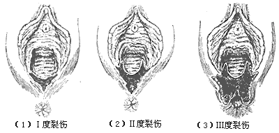
**Figure 1** Perineal laceration
3. **Placental factors**: Incomplete placental separation or retained placenta in the uterine cavity may present clinically as uterine atony, with the placenta failing to be expelled and significant bleeding. A constriction ring may be observed in the lower uterine segment in cases of placental incarceration. Partial placental adhesion to the uterine wall can lead to incomplete separation, and the retained placenta may impair uterine contractions, leaving the placental site blood sinuses open and bleeding. In cases of complete adhesion, the placenta fails to separate and expel on time, and diagnosis is confirmed only when manual removal reveals the placenta firmly attached to the uterine wall. Partial placental implantation may result in persistent bleeding from the non-implanted portion, often confused with placental adhesion. Manual removal may reveal the placenta partially or entirely fused with the uterine wall, making separation difficult. Placental remnants are typically diagnosed during routine post-expulsion examination of the placenta and membranes, where defects in the placental surface or torn blood vessels at the membrane edges indicate retained placental tissue or accessory placenta.
4. **Coagulation disorders**: A tendency toward bleeding before or during pregnancy, combined with placental separation or birth canal injury, may lead to coagulation dysfunction and excessive bleeding.
bubble_chart Treatment Measures
The principle is to quickly stop bleeding, correct hemorrhagic shock, and control infection.
1. Uterine atony: Strengthening uterine contractions is the fastest and most effective method to stop bleeding caused by uterine atony. The birth attendant quickly places one hand on the uterine fundus, with the thumb on the anterior wall and the other four fingers on the posterior wall, performing rhythmic and gentle massage on the uterine fundus. After massage, the uterus begins to contract (Figure 2). Alternatively, one hand can be placed in the anterior vaginal fornix to support the anterior uterine wall, while the other hand presses the posterior uterine wall from the abdomen, flexing the uterus anteriorly. Both hands then press and massage the uterus simultaneously (Figure 3). If necessary, another hand can be placed above the pubic symphysis to press the midline of the lower abdomen and push the uterus upward. Massaging the uterus must emphasize holding the uterine body with the hand, elevating it above the pelvic cavity, and performing rhythmic, gentle massage. The pressing time should continue until the uterus regains normal contractions and maintains a contracted state. During massage, 10U of oxytocin can be administered intramuscularly or slowly intravenously (added to 20ml of 10-25% glucose solution), followed by intramuscular or intravenous injection of 0.2mg ergonovine (use with caution in patients with heart disease). Then, 10-30U of oxytocin can be added to 500ml of 10% glucose solution for intravenous drip to maintain good uterine contraction.
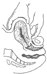
Figure 2: Abdominal massage of the uterine fundus
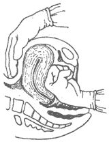
Figure 3: Abdominal-vaginal bimanual uterine massage method
The above measures often lead to uterine contraction and rapid hemostasis. If they are ineffective, the following measures can be taken:
(1) Uterine packing: In modern obstetrics, gauze packing of the uterine cavity is rarely used to treat uterine bleeding. If this procedure is necessary, it should be performed early, as delayed intervention often yields poor results due to weakened uterine muscle contraction. The method involves sterilizing the area, with the operator fixing the uterine fundus with one hand on the abdomen and using the other hand or an ovum forceps to insert a 2cm-wide gauze strip into the uterine cavity. The gauze must be packed tightly from the fundus outward (Figure 4). After packing, bleeding usually stops, and the patient's condition improves with anti-shock treatment. If sausage-shaped gauze wrapped with absorbent cotton is used instead of a gauze strip, the effect may be better. The gauze strip should be slowly removed after 24 hours, preceded by intramuscular injection of oxytocin or ergonovine to induce uterine contraction. After uterine packing, the patient's general condition, blood pressure, pulse, and other vital signs should be closely monitored. Attention should be paid to changes in uterine fundus height and size to avoid the false impression of hemostasis if the gauze is loosely packed only in the lower uterine segment, allowing continued bleeding in the uterine cavity without visible vaginal bleeding.
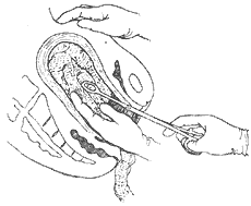
Figure 4: Method of uterine cavity gauze packing
(2) Ligation of uterine arteries: If massage fails or uterine contraction does not resume after half an hour of massage, bilateral ascending branch ligation of the uterine arteries via the vagina can be performed. After sterilization, two long rat-tooth forceps are used to clamp the anterior and posterior lips of the cervix and gently pull downward. The lateral walls of the cervix at the upper end of the vaginal portion are sutured with No. 2 catgut, penetrating about 0.5cm into the tissue. If ineffective, laparotomy should be performed promptly to ligate the ascending branches of the uterine arteries. This involves suturing the lateral cervical wall at the level of the internal cervical os, about 1cm from the lateral cervical wall, ensuring the ureter is not involved. The suture penetrates about 1cm into the cervical tissue, and the same procedure is performed on both sides. Uterine contraction indicates effectiveness.
(3) Ligation of the internal iliac stirred pulse: If the above measures are still ineffective, the starting points of the bilateral internal iliac stirred pulse can be isolated and ligated with No. 7 silk thread. After ligation, the uterus is generally seen to contract well. This measure can preserve the uterus and fertility, and is easy to perform during cesarean section.
(4) Uterus removal: When ligating blood vessels or packing the uterine cavity remains ineffective, a subtotal hysterectomy should be performed immediately without hesitation to avoid delaying the rescue opportunity.
2. Laceration of the birth canal: The effective measure for hemostasis is timely and accurate repair and suturing.
Generally, severe cervical lacerations may extend to the fornix and even into adjacent tissues. If a cervical laceration is suspected, the cervix should be exposed under disinfection. Using two oval forceps, clamp the anterior lip of the cervix side by side and pull toward the vaginal opening. Gradually move the forceps clockwise to observe the cervix under direct vision. If a laceration is found, suture it with catgut. The first stitch should start slightly above the apex of the laceration, and the last stitch should end 0.5 cm from the outer edge of the cervix. Suturing to the outer edge may lead to cervical stenosis in the future (Figure 5).
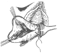
Figure 5: Suturing of cervical laceration
When suturing vaginal lacerations, ensure the suture reaches the base to avoid leaving dead space. The suturing should achieve good tissue alignment and hemostasis. During vaginal suturing, avoid stitching through the rectum. Suturing perpendicular to the direction of blood vessels can enhance hemostasis.
Perineal lacerations should be sutured according to anatomical layers, including the muscle layer and submucosal layer, followed by the vaginal mucosa and perineal skin.
3. Placental factors: The key to treatment is early diagnosis and prompt removal of the causative factor.
Incomplete placental separation, retention, or adhesion can be manually removed. For residual tissue that cannot be manually extracted, a large curette may be used. If manual separation of the placenta reveals unclear attachment boundaries, avoid forcibly separating the placenta with fingers, as this may indicate placenta accreta. In such cases, a laparotomy and uterine incision should be performed for examination. If confirmed, a subtotal hysterectomy is advisable. For a placenta trapped above a uterine constriction ring, ether anesthesia should be administered to relax the constriction ring before manual removal of the placenta.
4. Coagulation disorders: If detected early in pregnancy, an induced late abortion should be performed as soon as possible with the cooperation of an internist. If discovered in the mid or advanced stages of pregnancy, active treatment should be pursued with an internist to eliminate the cause or significantly improve the condition. During childbirth, while treating the underlying cause, address even minor bleeding promptly by using medications to improve coagulation, transfusing fresh blood, and preparing for anti-shock and acidosis correction measures. Refer to relevant sections for details.
For postpartum metrorrhagia, while controlling bleeding, actively manage hemorrhagic shock to rapidly improve the patient's condition. Use antibiotics to control infection.
Preventing postpartum metrorrhagia can significantly reduce its incidence. Preventive measures should be implemented throughout the following stages.
1. Ensure proper pre-pregnancy and prenatal care. Begin prenatal check-ups and monitoring early in pregnancy, and terminate the pregnancy promptly in the first trimester for those who are not suitable for pregnancy.
2. Prepare for early intervention in women at high risk of postpartum metrorrhagia. These include: ① women with multiple pregnancies, multiple deliveries, or a history of multiple uterine surgeries; ② older primiparas or very young pregnant women; ③ those with a history of uterine myomectomy; ④ genital hypoplasia or malformation; ⑤ pregnancy-induced hypertension; ⑥ those with diabetes, hematologic diseases, etc.; ⑦ prolonged labor due to uterine inertia; ⑧ those who undergo assisted delivery with vacuum extraction or forceps, especially when combined with uterotonics; ⑨ cases of fetal demise, etc.
3. Closely monitor the mother during the first stage of labor, ensuring adequate hydration and nutrition to avoid excessive fatigue. Administer pethidine intramuscularly if necessary to allow the mother to rest.
4. Pay attention to the management of the second stage of labor, guiding the mother to use abdominal pressure appropriately and correctly. For those at risk of postpartum metrorrhagia, a highly skilled physician should be present. Perform episiotomy or median perineal incision when indicated. Standardize delivery techniques to ensure smooth delivery of the fetal head, shoulders, and body. For cases of uterine inertia, administer 10U of oxytocin intramuscularly after the delivery of the shoulders, followed by intravenous oxytocin to enhance uterine contractions and reduce bleeding.
5. Properly manage the third stage of labor, accurately collecting and measuring postpartum metrorrhagia. After signs of placental separation appear, gently press the lower uterine segment and lightly pull the umbilical cord to assist in the complete expulsion of the placenta and membranes, and carefully inspect for completeness. Examine the soft birth canal for lacerations or hematomas. Assess uterine contractions and perform uterine massage to promote contraction.
6. After placental delivery, the mother should remain in the delivery room for observation for 2 hours, as approximately 80% of postpartum metrorrhagia occurs within this period. Focus on monitoring general condition, vital signs, vaginal bleeding, and uterine contractions. However, do not overlook bleeding after 12 hours. Provide the mother with necessary instructions, and ensure regular check-ups by medical staff to address any issues promptly.
7. For those with significant blood loss but no signs of shock, initiate blood volume replacement early, as it is far more effective than waiting until shock occurs to administer the same volume.
8. Early breastfeeding stimulates uterine contractions and reduces vaginal bleeding.


