| disease | Inguinal Sliding Hernia |
A sliding inguinal hernia refers to a hernia where the protruding organ and/or its mesentery form part of the hernia sac. On the right side, the commonly herniated organ is the cecum, while on the left side, it is the sigmoid colon. The serosal layer of the cecum or sigmoid colon forms the posterior wall of the hernia sac and folds back on both sides to form the lateral and anterior walls of the sac. Occasionally, the herniated organ may be the bladder.
bubble_chart Clinical Manifestations
Sliding hernias are relatively uncommon and lack distinctive clinical manifestations, making preoperative diagnosis difficult. However, due to their structural characteristics, sliding hernias are often irreducible, typically presenting as irreducible hernias. In elderly, obese patients or those with a long history of hernias, if the hernia mass is difficult to reduce or only partially reducible, the possibility of a sliding hernia should be considered.
bubble_chart Treatment Measures
Surgical treatment should be performed in all cases. The key points of the surgery are to first free and reduce the herniated organs, reconstruct a complete hernial sac, and then perform high ligation and hernia repair.
1. Hernial sac formation and high ligation
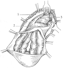
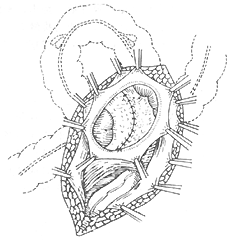
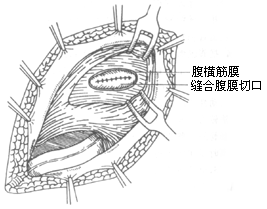
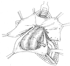
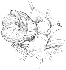
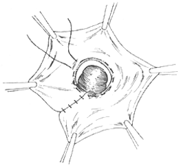
(1) Bevan method (Figure 1): This is a commonly used method but is only suitable for smaller sliding hernias, such as cecal herniation. The specific steps are as follows: after incising the hernial sac, make an arc-shaped incision along the edge of the cecum 2 cm away, cutting the peritoneum. The ends of the incision must reach the neck of the hernial sac to ensure that a complete hernial sac can be formed for high ligation. Carefully free the cecum to the level of the internal ring, avoiding accidental injury to the mesenteric vessels and spermatic cord vessels. At this point, the herniated cecum can be reduced, and the two ends of the arc-shaped peritoneal incision can be approximated and sutured longitudinally to form a complete hernial sac for high ligation.
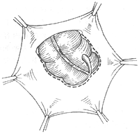 | 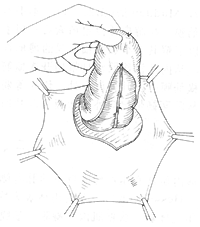 |
(1) Make an arc-shaped incision in the peritoneum 1–2 cm from the edge of the cecum | (2) Free the posterior wall of the cecum to the level of the internal ring |
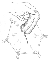 | 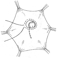 |
(3) Suture the arc-shaped peritoneal incision longitudinally | (4) Reduce the cecum and perform high ligation of the hernial sac |
Figure 1 Bevan surgery
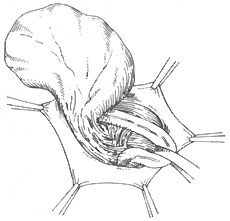
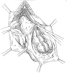
(2) La Roque method (Figure 2): Suitable for larger sliding hernias, such as longer herniated intestines like the sigmoid colon. This method is more reliable. The specific steps are as follows: incise the anterior wall of the hernial sac and free the posterior side of the intestine up to the internal ring. If the herniated intestine is long, care must be taken during freeing to avoid accidental injury to its mesenteric vessels. Then, 3 cm above the internal ring, separate the internal oblique muscle and transverse abdominis muscle along the direction of the muscle fibers, taking care not to injure the iliohypogastric nerve. Incise the peritoneum, reduce the freed herniated intestine through the internal ring, and bring it out through the abdominal incision. The freed posterior surface of the original intestine is then flipped to the front, and the parietal peritoneum between the hernial sac incision and the peritoneal incision is also everted. Trim the excess hernial sac so that the remaining edges can be approximated and sutured to cover the freed surface of the intestine, forming the serosal layer behind the mesentery. Reduce the intestine and finally suture the peritoneal incision.
 | 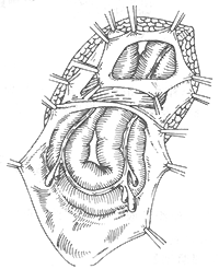 |
(1) The dotted line indicates the incision of the transversus abdominis membrane, cutting through the anterior wall of the hernial sac. 1. External oblique aponeurosis membrane 2. Internal oblique muscle 3. Transversus abdominis membrane | (2) Incise the abdominal membrane approximately 3cm above the internal ring |
 |  |
(3) The freed and slipped intestine is pulled out through the internal ring from the abdominal incision | (4) The freed surface of the intestine is turned to the front and serosalized |
 |  |
(5) Reduce the intestine | (6) Suture |
Figure 2 La Roque surgery
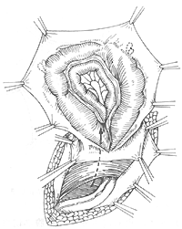
Additionally, Ponka introduced a surgical method suitable for larger sliding hernias (Figure 3): First, separate the hernial sac along with the slipped intestine from the spermatic cord to the deep surface of the internal ring, taking care not to injure the mesenteric membrane of the intestine or the spermatic vessels. Incise the anterior wall of the hernial sac and cut along both sides of the intestine to the deep surface of the internal ring. Then, suture the two incised edges behind the freed surface of the intestine to form a complete internal ring, reduce the intestine, and perform high ligation of the hernial sac. This surgery does not require serosalization of the freed surface behind the intestine.
 |  |
| (1) Dissect the hernial sac to the internal ring | (2) Incise the anterior wall of the hernial sac |
 |  |
| (3) Cut the abdominal membrane along both sides of the intestine to the internal ring | (4) Suture the two incised edges of the abdominal membrane behind the freed surface of the intestine to form a complete internal ring |
 | |
| (5) Reduce the intestine and perform high ligation of the hernia sac | |
Figure 3 Ponka's operation
2. Herniorrhaphy The characteristic of sliding hernia is the enlargement of the hernia ring, with severe impairment of the strength of the abdominal wall tendon membrane and transversus abdominis membrane. Therefore, the Bassini, Halsted, or McVay methods are more commonly used.


