| disease | Carcinoma of Endometrium |
| alias | Uterine Corpus Cancer, Carcinoma of the Endometrium |
Carcinoma of the endometrium (carcinoma of the endometrium), also known as carcinoma of the corpus uteri, is a common malignant tumor in gynecology, second only to cervical cancer.
bubble_chart Etiology
The true cause of carcinoma of endometrium remains unknown to date, but its risk factors have long been noted. The risk factors include:
1. **Obesity** — Excessive fat increases the storage of estrogen and the conversion of androstenedione to estrone in plasma. This increase in free, active estrone may act as a carcinogenic or promoting factor for carcinoma of endometrium.
2. **Diabetes** — Patients with diabetes or abnormal glucose tolerance have a 2.8 times higher risk of developing carcinoma of endometrium compared to normal individuals.
3. **Hypertension** — Endometrial carcinoma is often accompanied by hypertension.
The coexistence of obesity, diabetes, and hypertension in patients with carcinoma of endometrium is referred to as the "triad of endometrial carcinoma" or "endometrial carcinoma syndrome." These three factors may be related to a high-fat diet, which has a direct association with carcinoma of endometrium.
4. **Menstrual disorders** — Women with endometrial carcinoma have a threefold higher incidence of menstrual irregularities and heavy bleeding compared to normal women.
5. **Early menarche and delayed menopause** — Women who experience menarche before the age of 12 have a 60% higher incidence of endometrial carcinoma than those who experience it after 12. The age of menopause in endometrial carcinoma patients is delayed by six years compared to normal women.
6. **Parity** — Endometrial carcinoma is more common in women with multiple pregnancies, nulliparity, or infertility.
7. **Polycystic ovary syndrome (PCOS)** — Characterized by anovulation, which exposes the endometrium to high, continuous estrogen levels without progesterone regulation or cyclical endometrial shedding, leading to hyperplastic changes.8. **Ovarian tumors** — Estrogen-secreting tumors such as granulosa cell tumors and theca cell tumors can cause menstrual irregularities, postmenopausal bleeding, endometrial hyperplasia, and endometrial carcinoma.
9. **Atypical endometrial hyperplasia** — This may represent a stage in the development of endometrial carcinoma or occur independently. Grade III atypical hyperplasia can be considered as carcinoma in situ of the endometrium.
10. **Exogenous estrogen** — Women taking estrogen have a significantly increased risk of developing carcinoma of endometrium. The risk depends on the dose, duration of use, whether progesterone is combined, whether treatment is interrupted, and individual patient characteristics. The risk decreases after discontinuation but may persist for several years. There is now substantial evidence supporting a causal relationship between estrogen and endometrial carcinoma.
Among estrogens, estriol (E3) does not promote endometrial hyperplasia, whereas E2, E1, ethinyl estradiol, or conjugated estrogens readily induce endometrial hyperplasia, increasing the risk of carcinoma of endometrium.
bubble_chart Pathological Changes
I. Gross Pathology: Can be divided into three types: diffuse, localized, and polypoid.
(1) Diffuse Type: The lesion may involve all or most of the endometrium. In its early stages, it is difficult to distinguish from hyperplastic endometrium. However, upon careful examination, the cancerous area still exhibits some characteristics, with identifiable boundaries between it and the normal endometrium. The mucous membrane of the cancer is thickened, rough, and has irregular polypoid projections, whereas benign endometrial hyperplasia is softer with a smooth surface. Malignant polypoid projections are larger, hard, brittle, and may have superficial ulcers. In advanced stages, the lesion shows ulceration and necrosis, involving the entire endometrium. In rare cases, it may even spread and invade the endocervical canal or extend to the vaginal fornix. In contrast, benign polypoid endometrial hyperplasia is confined to the area above the internal cervical os, as the cervix does not respond to the abnormal endocrine function causing this endometrial hyperplasia (Photo 1).
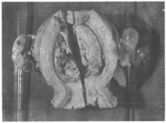
Photo 1: A cross-section of uterine corpus carcinoma, showing the tumor located on the posterior uterine wall with infected necrosis.
In addition to spreading within the endometrium, the cancer may invade the myometrium at a certain stage, even penetrating the uterine serosa and metastasizing to the ovaries, parametrium, rectum, and bladder. In advanced stages, the tumor surface undergoes necrosis and ulceration, often with secondary infection.
(2) Localized Type: Less common. The cancer is confined to a portion of the endometrium, with an appearance similar to the diffuse type. The superficial cancerous area may not be extensive, but it deeply invades the myometrium, causing uterine enlargement or necrotic infection leading to uterine wall ulceration or even perforation. Advanced stages similarly involve surrounding erosion or metastasis.
The localized type may present as polypoid, cauliflower-like, or nodular. The former is more common in early cases, while the latter is often seen in advanced cases, usually accompanied by myometrial invasion. Localized tumors are mostly located at the fundus or cornua of the uterus. Polypoid carcinoma closely resembles ordinary benign endometrial polyps but differs from the soft, smooth-surfaced benign endometrial polyps. The polypoid projections of the cancer are larger, brittle, and often show surface necrosis. Sometimes, polypoid carcinomas are small but entirely composed of malignant tissue and have already deeply invaded or penetrated the myometrium. Occasionally, polypoid carcinomas are few in number and may be entirely removed during diagnostic curettage, leaving no trace of the tumor in the resected uterine specimen. Of course, the possibility of specimen mislabeling during curettage should also be considered, and verification should be performed if suspicious to avoid misdiagnosing a true patient.
Polypoid endometrial carcinoma often occurs in the uterine cornua and is commonly seen in postmenopausal women.II. Microscopic Examination: Four types are introduced:
(1) Adenocarcinoma: Accounts for about 80–90%. Microscopically, endometrial glands are increased in number, vary in size, and are disorganized, showing a prominent "back-to-back" pattern. The epithelium may sometimes be papillary, protruding into the uterine cavity to form secondary glands, creating an "adenoma within adenoma" appearance. The cancer cells are large, irregular, with polymorphic and hyperchromatic nuclei, scant cytoplasm, and frequent mitotic figures. The stroma is sparse with inflammatory cell infiltration. Poorly differentiated adenocarcinomas show fewer glands, loss of structure, and formation of solid cancerous masses.
The International Federation of Gynecology and Obstetrics (FIGO, 1970) proposed a three-tier histological classification for endometrial carcinoma: - Grade I (Well-differentiated carcinoma): Usually confined to the endometrium, occasionally showing single or stratified papillary epithelium with irregular arrangement and scant stroma. - Grade II (Moderately differentiated carcinoma): Slightly poorer differentiation, with less distinct glandular outlines and partial solid cancerous masses. Cells lose polarity, and mitotic figures are common. - Grade III (Poorly differentiated or undifferentiated carcinoma): Extremely poor differentiation, with loss of glandular structure and predominantly solid cancerous masses.
(2) Adenoacanthoma: Also known as adenosquamous carcinoma. Microscopically, it is characterized by adenocarcinoma containing clusters of well-differentiated benign squamous epithelium, with visible intercellular bridges and keratinization patterns or the formation of keratin pearls.
(3) Adeno-squamous carcinoma: Also known as mixed carcinoma, the tumor tissue contains both adenocarcinoma and squamous cell carcinoma components.
(4) Clear cell carcinoma: The tumor exhibits a tubular structure, with microscopy revealing numerous small tubes of varying sizes arranged back-to-back, lined with transparent hobnail-like cells. These cells are characterized by scanty cytoplasm and large nuclei protruding into the lumen, with collagen fibers present in the stroma.
bubble_chart Clinical Manifestations
1. Symptoms Early-stage patients may show no obvious symptoms and are often discovered incidentally during routine screenings or gynecological examinations for other reasons. Once symptoms appear, they typically manifest as:
(1) Uterine bleeding: Irregular vaginal bleeding around menopause is the primary symptom of carcinoma of the endometrium. The bleeding is usually light to moderate, rarely heavy. Younger or perimenopausal patients may easily mistake it for menstrual irregularities and delay seeking medical attention. Even doctors often overlook it. Some patients may experience delayed menstrual cycles, but the pattern is irregular. Postmenopausal patients usually present with persistent or intermittent vaginal bleeding. Endometrial carcinoma patients generally do not experience contact bleeding. In advanced stages, the bleeding may contain necrotic tissue fragments.
(2) Vaginal discharge: Since adenocarcinoma grows within the uterine cavity, the chance of infection is lower compared to cervical carcinoma. In the initial stage [first stage], there may only be a small amount of bloody leucorrhea. However, if infection and necrosis occur later, a large amount of foul-smelling, pus-like discharge may be expelled. Sometimes, the discharge may contain small fragments of cancerous tissue. If the uterine cavity accumulates pus, it can cause fever, abdominal pain, and leukocytosis, leading to rapid deterioration of the patient's general condition.
(3) Pain: Due to the accumulation of cancerous tissue, blood, and discharge, irregular uterine contractions may cause paroxysmal pain, occurring in about 10–46% of patients. This symptom is more common in advanced stages. If the cancer penetrates the serous membrane or invades surrounding connective tissue, the bladder, rectum, or compresses other structures, it can also cause pain, often persistent and progressively worsening. The pain may radiate from the lower back or abdomen to the thighs and knees.
(4) Other symptoms: In advanced stages, patients may palpate an enlarged uterus or adjacent organs in the lower abdomen, leading to swelling and pain in the legs or compression of the ureter, resulting in hydronephrosis or kidney atrophy. Systemic manifestations such as anemia, weight loss, fever, and cachexia may also occur.
Endometrial carcinoma typically occurs at a later age, making concurrent pregnancy unlikely. However, there have been rare case reports of coexisting pregnancy or tubal pregnancy in the literature.
2. Signs
(1) Systemic manifestations: A significant proportion of patients have diabetes, hypertension, or obesity. Anemia may occur in patients with prolonged bleeding. In advanced stages, due to cancer-related wasting, pain, loss of appetite, fever, etc., cachexia may develop.
(2) Gynecological examination findings: In early stages, the pelvic reproductive organs often show no significant abnormalities, with the uterus appearing normal in about 40% of cases. If fibroids are present or the disease progresses to an advanced stage, the uterus may enlarge. Postmenopausal women with a uterus that is not atrophic but instead full and firm should be particularly vigilant. The ovaries may appear normal, enlarged, or possibly associated with feminizing tumors. If palpation is unclear due to obesity, pain, or lack of cooperation during bimanual examination, it is unnecessary to insist on a definitive assessment, as diagnostic criteria do not rely solely on uterine size. The cervix usually shows no visible abnormalities unless the cancer has invaded it in advanced stages, where cancerous tissue may protrude from the cervical os. Parametrial infiltration is a result of cervical involvement.
(3) Metastatic lesions: Advanced-stage patients may have enlarged, hardened, or fused lymph nodes in the groin or signs of metastasis to the lungs, liver, or other organs.
bubble_chart Auxiliary Examination
The diagnostic rate of vaginal cytology for endometrial carcinoma is lower than that for cervical cancer, due to the following reasons: ① Columnar epithelial cells do not shed frequently; ② When shed cells pass through the cervical canal and reach the vagina, they often undergo dissolution and degeneration, making them difficult to identify; ③ Sometimes the cervical canal is narrow or obstructed, preventing shed cells from reaching the vagina. To improve the positive diagnostic rate, many scholars have refined the sampling sites and methods, and with advancements in diagnostic techniques, the positive diagnostic rate for endometrial carcinoma has significantly increased.
For cytological examination of endometrial carcinoma, samples taken from the uterine cavity can greatly enhance the positive rate, often reaching around 96%, which is not lower than the positive rate of cervical scrapings for cervical carcinoma.
1. **Ultrasound Examination** Ultrasound examination of the uterus is significant for assessing the size and location of endometrial carcinoma in the uterine cavity, the extent of myometrial invasion, whether the tumor has penetrated the uterine serosa or involves the cervical canal, etc. The diagnostic accuracy rate ranges from 79.3% to 81.82%. It has been reported that for patients over 45 years old, when compared with hysteroscopy and biopsy, the accuracy of ultrasound is approximately 87%. Additionally, Xie Yanggui et al. performed ultrasound examinations using the UICC staging method, correlating tumor location, myometrial invasion, parametrial involvement, and adjacent organ involvement with surgical exploration and pathology, achieving a staging accuracy rate of 92.9%. Ultrasound is non-invasive and free from radioactive harm, making it a routine examination for endometrial carcinoma. It is particularly valuable for assessing myometrial invasion and clinical staging.
2. **Diagnostic Curettage** Curettage is an indispensable method for definitive diagnosis. It is essential not only to confirm the presence of cancer but also to determine its location. Misdiagnosing cervical adenocarcinoma as endometrial carcinoma and proceeding with a standard hysterectomy is clearly inappropriate; similarly, mistaking endometrial carcinoma for cervical adenocarcinoma is also inadvisable. However, microscopic examination alone cannot distinguish between cervical adenocarcinoma and endometrial carcinoma. Therefore, fractional curettage is necessary. First, a small curette is used to sample tissue from the cervical canal, followed by sampling from the uterine cavity, including the cornual regions and the anterior and posterior walls of the uterine body. The samples are separately labeled and sent for pathological examination. If resistance is encountered at the internal os, the cervix may be slightly dilated to size 5. During fractional curettage, errors may occur, such as mistaking uterine cavity contents for cervical canal cancer, or misidentifying endometrial carcinoma prolapsing into the cervical canal as cervical canal cancer or uterine body cancer involving the cervical canal. Alternatively, excessive cervical canal cancer tissue may be inadvertently introduced into the uterine cavity by the curette, leading to a false impression of cervical carcinoma extending into the uterine cavity. Such scenarios indicate advanced disease and should be managed with surgical approaches appropriate for cervical carcinoma.
During curettage, the force applied should be appropriate. If cancer tissue is clearly visible after one or two passes, further scraping should be avoided to prevent uterine perforation or artificial spread of cancer or inflammation. If no obvious cancer tissue is obtained, a thorough uterine cavity scraping is necessary, with attention to the fundus and cornual regions. All scraped tissue should be sent for pathological examination to confirm or exclude early endometrial carcinoma. If genital tract inflammation is present, the procedure should be delayed until the inflammation is controlled. Alternatively, a specialized fine metal tube (endometrial suction curette) can be used to minimize uterine wall contact, but standard suction tubes for late-term abortions should not be used, and suction pressure should not be excessive. Generally, a syringe can be attached for gentle suction to avoid excessive tissue injury. If suction yields minimal tissue, conventional curettage may still be performed. If the diagnosis remains uncertain despite clinical suspicion, regular follow-up examinations are recommended.
The accuracy of endometrial biopsy ranges from 87% to 100%. Its advantage lies in providing a histological diagnosis, which is definitive. However, its drawback is the potential for blind or insufficient sampling, particularly in postmenopausal patients. Hence, there is a growing preference for direct biopsy under hysteroscopic guidance.
It is worth noting that according to the new FIGO clinical staging, fractional diagnostic curettage is no longer applicable. Moreover, literature reports indicate that fractional curettage may lead to confusion between cervical canal and endometrial tissues, resulting in misjudgment; or early cervical involvement may be overlooked during curettage. Additionally, cases with lower uterine segment involvement exhibit similar lymphatic metastasis patterns to those with cervical involvement, casting doubt on its value. However, under the current practical circumstances in China, fractional diagnostic curettage remains an indispensable primary diagnostic method. Its diagnostic accuracy rate is as high as 94–97.5%, and the procedure is simple and safe. Of course, since it is performed non-visually, there is still a possibility of occasionally missing fistula disease. Therefore, a negative curettage result cannot completely rule out the presence of cancer.
III. Hysteroscopy Due to the application of fiber-optic light sources and improvements in uterine distension media, this long-stagnant technique has seen renewed development in recent years. CO2 gas for uterine distension provides a clear field of view and is very safe when used with a flowmeter device. Hysteroscopy allows not only observation of the uterine cavity but also the cervical canal, especially with the use of microhysteroscopy, enabling more detailed examination. The recently developed contact hysteroscope eliminates the need for uterine distension, making the procedure simpler and safer. Hysteroscopy can assess the location, size, and boundaries of tumors—whether localized or diffuse, exophytic or endophytic—and whether the cervical canal is involved. Biopsies of suspicious lesions aid in detecting smaller or early-stage abnormalities. The diagnostic accuracy of hysteroscopy is 94% for endometrial cancer and 92% for endometrial intraepithelial neoplasia. Direct biopsy increases accuracy to 100%. Care must be taken during the procedure to prevent complications such as bleeding, infection, and perforation.
The morphological features of carcinoma of endometrium under hysteroscopy are detailed in Section 6 of Chapter 12.
IV. Retroperitoneal Lymphangiography This technique clarifies whether metastases are present in the pelvic and para-aortic lymph nodes, aiding in treatment planning. For stages I and II, the rates of pelvic lymph node metastasis are 10.6% and 36.5%, respectively.
V. Computed Tomography (CT) and Magnetic Resonance Imaging (MRI) CT has certain diagnostic value for endometrial cancer, providing clear images and accurate delineation of tissue microstructures. It can precisely measure tumor size and extent, with 83% accuracy in determining the stage of localized uterine wall tumors. CT also identifies tumor spread to surrounding connective tissue, pelvic and para-aortic lymph nodes, pelvic walls, and peritoneal metastases. It is particularly superior to ultrasound for examining obese women. MRI, with its three-dimensional scanning (superior to CT's two-dimensional scanning), can depict stage a endometrial cancer and the extent of myometrial invasion, showing irregular high-signal endometrial thickening and the disappearance of the low-signal junctional zone between the endometrium and myometrium. The overall diagnostic accuracy of MRI is 88%, and it accurately assesses myometrial invasion (except post-radiotherapy), enabling more precise tumor staging. However, MRI is less ideal for detecting smaller pelvic metastases or lymph node involvement.
CT and MRI offer unique advantages in diagnosing endometrial cancer, but their accuracy is not significantly higher than that of B-ultrasound, and they are more expensive, increasing the financial burden on patients. Generally, most patients can achieve a definitive diagnosis through cytology, B-ultrasound, and subsequent diagnostic curettage with pathological examination.
In addition to a detailed medical history, symptoms, and signs, the final diagnosis must be based on the histopathological examination of the endometrial tissue.
1. **Medical History** Patients with endometrial carcinoma are mostly elderly women, often with delayed menopause or irregular menstruation. They are frequently infertile or have few childbirths and may also have obesity, hypertension, or diabetes. Postmenopausal women with irregular vaginal bleeding or foul-smelling discharge should be particularly monitored. For younger patients with irregular vaginal bleeding, the cause should be carefully investigated, especially if treatment is ineffective, and diagnostic curettage should be performed. Vaginal discharge and abdominal pain are symptoms of advanced-stage disease.
2. **Clinical Examination** In the early stages, routine gynecological examinations often reveal no abnormalities—the uterus is not enlarged, the cervix is smooth, and the adnexa are normal. In advanced stages, the uterus may be larger than expected for the patient’s age, and bloody leucorrhea or necrotic cancerous tissue may be found on the examining glove after bimanual palpation. In some cases, polypoid masses may protrude from the cervical os. However, endometrial carcinoma can coexist with uterine fibroids, so an enlarged uterus does not necessarily indicate advanced-stage endometrial carcinoma.
3. **Cytological Examination** The diagnostic accuracy of vaginal cytology for endometrial carcinoma is lower than that for cervical cancer due to: - Columnar epithelial cells not shedding frequently. - Shed cells often degrade or dissolve before reaching the vagina. - Narrow or obstructed cervical canals may prevent cells from reaching the vagina. To improve diagnostic accuracy, researchers have refined sampling methods and techniques, significantly increasing the positive detection rate for endometrial carcinoma.
For example, a study of 103 endometrial carcinoma cases compared vaginal smears with uterine cavity aspiration (to avoid contamination from cervical or vaginal cells). The positive rate was 74.5% for vaginal smears and 93% for uterine cavity aspirates, while diagnostic curettage had a 98% positive rate. Two cases were negative on curettage but positive on aspiration, suggesting that combining both methods can enhance detection rates, potentially reaching 100%. The cytological positivity rate also correlates with histological types, being slightly higher for well-differentiated adenocarcinomas and adenocanthomas. Improved uterine cavity aspiration devices can collect tissue fragments for analysis, offering a simple alternative for patients contraindicated for curettage. The cellular morphology in uterine aspirates differs from vaginal smears, featuring not only round cancer cells but also giant cells 5–6 times larger than normal, with nuclei 40–50 μm in diameter (normal: 5–7 μm). These cells exhibit polymorphism, multinucleation, hyper- or hypochromasia, and naked nuclei.
For the cytological examination of carcinoma of endometrium, specimens taken from the uterine cavity can significantly increase the positive rate, often reaching about 96%, which is not lower than the positive rate of cervical scrapings for cervical carcinoma. There are many methods for specimen collection domestically and internationally, such as the intrauterine membrane aspiration method using a 3mm metal tube connected to a syringe for suction; the spiral device method (composed of a soft plastic spiral scoop and a paddle-shaped scraper) for specimen collection; uterine cavity irrigation; the uterine membrane brush method; the sponge biopsy method (using a V-shaped polypropylene sponge, 5mm at the base with a thread attached to the top, inserted into the uterine cavity with an IUD inserter, and pulled out after the sponge absorbs tissue); and the uterine cavity scanner. Domestically, Zhou Weixu found the self-made plastic tube method for uterine cavity aspiration smears to be of certain value. The author used a nylon micro-brush (initially modified from discarded bronchoscope micro-brushes, now successfully trial-produced domestically) to sweep the cervical canal and uterine cavity separately. The one-time sweep positive rate for carcinoma of endometrium in the uterine cavity was 96% (24/25), while in the cervical canal it was 72% (18/25). If cases with cervical canal cancer involvement were excluded, the positive rate dropped to only 50%. This shows that for suspected endometrial cancer, direct smears from the uterine cavity are most ideal. This micro-nylon brush, due to its certain hardness and elasticity, can directly collect fresh cells from the endometrial or tumor surface and retain them in the brush. The procedure is similar to a curettage. The brush is swept up, down, left, and right four or five times before removal. After removal, the brush is directly smeared onto a glass slide. The operation and smear method are extremely simple, and the small brush easily enters the uterine cavity. It is a safe, simple, highly accurate diagnostic tool, and an easily promotable uterine cavity cell collector.
IV. B-ultrasound Examination Ultrasound examination of the uterus holds certain significance in assessing the size and location of the uterine cavity, the extent of myometrial invasion, whether the tumor has penetrated the uterine serosa or involved the cervical canal, etc., with a diagnostic accuracy rate of 79.3–81.82%. Reports indicate that for patients over 45 years old, when compared with hysteroscopy and biopsy, the accuracy of ultrasound is approximately 87%. Additionally, Xie Yanggui et al. conducted B-ultrasound examinations using the UICC staging method, correlating tumor location, myometrial invasion, parametrial involvement, and adjacent organ involvement with surgical exploration and pathology, achieving a staging accuracy rate of 92.9%. B-ultrasound is non-invasive and free from radioactive harm, making it one of the routine examinations for carcinoma of the endometrium. It is particularly valuable in assessing myometrial invasion and clinical staging.
V. Diagnostic Curettage Curettage is an indispensable method for definitive diagnosis. It is essential not only to confirm the presence of cancer but also to determine its location. Misdiagnosing cervical adenocarcinoma as carcinoma of the endometrium and proceeding with a standard hysterectomy would be inappropriate, just as mistaking carcinoma of the endometrium for cervical adenocarcinoma would be inadvisable. However, microscopic examination cannot distinguish between cervical adenocarcinoma and carcinoma of the endometrium. Therefore, fractional curettage is necessary. First, a small curette is used to collect tissue from the cervical canal, followed by entry into the uterine cavity to collect tissue from the cornua and the anterior and posterior walls of the uterine body. The samples are separately bottled, labeled, and sent for pathological examination. If resistance is encountered at the internal os, the cervix may be slightly dilated to a size 5. Fractional curettage often involves slight overextension into the cervical canal, leading to misidentification of uterine cavity contents as cervical canal cancer. Alternatively, carcinoma of the endometrium prolapsing into the cervical canal may be mistaken for cervical canal cancer or uterine body cancer involving the cervical canal. Conversely, excessive cervical canal cancer tissue may be inadvertently carried into the uterine cavity by the curette, leading to the false impression that cervical carcinoma has reached the uterine cavity. All these scenarios indicate advanced disease and should be managed with the surgical scope appropriate for cervical carcinoma.
During curettage, the force applied should be appropriate. If cancer tissue is clearly visible after one or two passes of the curette, further scraping should be avoided to prevent uterine perforation or artificial spread of cancer or inflammation. If no obvious cancer tissue is obtained, comprehensive uterine cavity scraping must be performed, with attention paid to the fundus and cornua. All collected tissue should be sent for pathological examination to confirm or exclude early carcinoma of the endometrium. If genital inflammation is present, the procedure should be postponed until the inflammation is controlled. Alternatively, a specialized fine metal tube (endometrial suction curette) may be used to minimize uterine wall contact, but the suction tubes currently used for late-term abortions should not be employed, and suction pressure should not be excessive. Generally, attaching a syringe for aspiration suffices to avoid excessive tissue injury. If suction yields minimal tissue, conventional curettage may still be performed. If the initial procedure is inconclusive but clinical suspicion remains, regular follow-up is recommended.
Endometrial biopsy has an accuracy rate of 87–100%. Its advantage lies in providing a histological diagnosis, which is definitive. However, its drawback is the potential for blind or insufficient sampling, particularly in postmenopausal patients where inadequate tissue is often obtained. Consequently, there is a growing trend toward direct biopsy under hysteroscopic observation.
It is worth noting that, according to the new FIGO clinical staging system, fractional diagnostic curettage is no longer applicable. Moreover, literature reports indicate that fractional curettage can lead to confusion between cervical canal and uterine cavity tissue, resulting in misdiagnosis. Early cervical involvement or fistula disease may also complicate the procedure. Additionally, lymph node metastasis in cases involving the lower uterine segment resembles that of cervical involvement, raising doubts about its value. However, given the current practical circumstances in China, fractional diagnostic curettage remains an indispensable primary diagnostic method, with a high accuracy rate of 94–97.5%. It is simple and safe to perform. Of course, as it is performed non-visually, there remains a slight possibility of missing fistula disease. Therefore, a negative curettage result does not entirely rule out the presence of cancer.
VI. Hysteroscopy Due to the application of fiber-optic light sources and improvements in uterine distension media, this long-stagnant technique has seen renewed development in recent years. CO2 gas for uterine distension provides a clear field of view and is very safe to use when equipped with a flowmeter device. Hysteroscopy not only allows observation of the uterine cavity but also the cervical canal, especially with the use of micro-hysteroscopy, enabling more detailed examination. The recently developed contact hysteroscope eliminates the need for uterine distension, making the procedure even simpler and safer. Under hysteroscopy, the location, size, and boundaries of cancerous lesions can be observed—whether they are localized or diffuse, exophytic or endophytic, and whether the cervical canal is involved. Biopsies of suspicious lesions can help detect smaller or early-stage abnormalities. The diagnostic accuracy of hysteroscopy is 94% for endometrial cancer and 92% for endometrial intraepithelial neoplasia. If direct biopsy is performed, the accuracy can reach 100%. During the procedure, care must be taken to prevent complications such as bleeding, infection, and perforation.
The morphology of carcinoma of endometrium under hysteroscopy is described in Section 6 of Chapter 12.
7. Retroperitoneal Lymphangiography This can clarify whether there is metastasis in the pelvic and para-aortic lymph nodes, aiding in the determination of treatment plans. For stages I and II, the pelvic lymph node positivity rates are 10.6% and 36.5%, respectively.
8. Computed Tomography (CT) and Magnetic Resonance Imaging (MRI) CT has certain diagnostic value for endometrial cancer. CT scans provide clear images and can accurately depict the fine structure of tissues, as well as precisely measure tumor size and extent. For tumors confined to the uterine wall, 83% can determine the stage of the disease. CT can also identify the spread of uterine tumors to surrounding connective tissues, pelvic and para-aortic lymph nodes, pelvic wall, and peritoneal metastatic nodules. It is particularly superior to ultrasound for examining obese women. MRI, being a three-dimensional scan (superior to CT's two-dimensional scan), can delineate stage a endometrial cancer. It can also depict the extent of tumor infiltration from the endometrium into the myometrium, manifested as irregular high-signal areas of thickened endometrium and the disappearance of the low-signal junctional zone between the endometrium and myometrium. The overall diagnostic accuracy of MRI is 88%, and it can accurately assess the degree of myometrial invasion (though not reliable post-radiotherapy), thereby providing a more precise estimation of tumor staging. However, MRI is less ideal for diagnosing smaller pelvic metastatic lesions and lymph node metastases.
CT and MRI have unique features in the diagnosis of endometrial cancer, but their diagnostic accuracy is not significantly higher than that of B-ultrasound. Moreover, they are expensive, increasing the financial burden on patients. Generally, most patients can achieve a definitive diagnosis through cytology, B-ultrasound, and subsequent diagnostic curettage with pathological examination.
bubble_chart Treatment Measures
The treatment principles for carcinoma of the endometrium should be comprehensively determined based on clinical staging, the degree of cancer cell differentiation, the patient's overall condition, and other factors. Since the majority of endometrial cancers are adenocarcinomas, which are not sensitive to radiotherapy, the primary treatment is surgery. Other treatments include radiotherapy, chemotherapy, and other medications as part of a comprehensive approach.
1. **Surgical Treatment** Bickenbach (1967) concluded that the efficacy of surgery alone is superior to radiotherapy alone, with a 5-year cure rate 20% higher for surgical treatment. According to a long-term follow-up study by Zhang Xiyin et al. in China involving 516 cases of endometrial cancer, the survival rate for surgery alone was 72%, while for preoperative radiotherapy plus surgery, it was 60%. The 5-, 10-, 15-, and 20-year survival rates were 85.9%–88.8%, 82.5%–85.8%, 81.4%–84.8%, and 77.3%–81.7%, respectively, demonstrating the effectiveness of surgical treatment. Surgery can clarify the extent of the lesion and accurately determine clinical staging, thereby guiding the appropriate surgical scope. Previously, according to the 1982 FIGO staging, Stage I cases typically underwent extrafascial total hysterectomy with bilateral salpingo-oophorectomy; Stage II cases underwent radical hysterectomy with bilateral pelvic lymphadenectomy. For Stages III and IV, surgery was performed whenever possible to resect the tumor and reduce its size, followed by adjuvant radiotherapy or progestin therapy. Otherwise, progestin therapy, radiotherapy, and/or chemotherapy were administered first to make surgery feasible, with additional adjuvant therapy post-surgery.
The 1988 FIGO clinical staging update advised clinicians that for Stage Ia cases, traditional extrafascial total hysterectomy with bilateral salpingo-oophorectomy and a 2 cm vaginal cuff is appropriate. For cases with myometrial invasion, especially deep invasion, the surgical scope should be expanded to radical hysterectomy with pelvic lymphadenectomy, as traditionally performed for Stage II. The para-aortic lymph nodes should be examined for enlargement, and if present, para-aortic lymph node biopsy or routine para-aortic lymphadenectomy should be performed. For Stages II and III, radical hysterectomy with pelvic and/or para-aortic lymphadenectomy should be performed as described. For Stage IV, debulking surgery should be attempted whenever possible. In 1972, Milton compared the 5-year survival rates between total hysterectomy and modified radical hysterectomy (without lymphadenectomy), finding 75.7% and 91.4%, respectively, suggesting that expanding the hysterectomy scope (at least modified radical hysterectomy) helps reduce postoperative recurrence rates. Additional considerations include:
(1) **Examination of ascites or peritoneal washings for cancer cells**: Upon opening the peritoneum, ascites should be collected and centrifuged to detect cancer cells. If no ascites is present, 200 ml of saline should be injected into the peritoneal cavity for washing, and the fluid centrifuged for analysis. If cancer cells are found (reported in 11.4% of Stage I cases, increasing significantly with higher tumor grades, e.g., 18.1% for Grade 3), adjuvant therapy should be added post-surgery.
(2) **Intraoperative assessment of myometrial invasion**: For Stage I cases with a uterus smaller than normal or when surgery time is limited, hysterectomy with salpingo-oophorectomy may be performed first, and the specimen examined for myometrial invasion. If gross examination is inconclusive, microscopic features to note include: ① Irregular, jagged glands in cancerous myometrial invasion, contrasting with round, smooth basal layer glands; ② Absence of endometrial stroma around invasive glands, unlike basal layer glands surrounded by stroma; ③ Significant edema around invasive foci.
If gross examination reveals cancer in the lower uterine segment, the surgical scope for Stage II should be followed.
(3) For those who have not undergone lymph node dissection: Routinely examine the lymph nodes in the pelvic and abdominal aorta regions. If enlarged lymph nodes are found, a biopsy should be performed at the very least. If technical conditions permit and the patient's condition allows, lymph node dissection may be performed.
II. Radiation Therapy Adenocarcinoma is not highly sensitive to radiotherapy, and the efficacy of radiation therapy alone is poor. However, for elderly patients or those with severe medical conditions who cannot undergo or are contraindicated for surgery, radiotherapy remains a treatment option with certain therapeutic benefits. Radiotherapy includes intracavitary and external beam irradiation. For intracavitary irradiation, 137Cs and 60Co are commonly used today, while radium has largely been abandoned. External beam irradiation often employs 60Co linear accelerators. According to reports by Wu Yuzhen et al. in China, intracavitary radiotherapy commonly uses the uterine packing method, with a low preoperative complication rate of 1%. External beam radiotherapy can be tailored to the primary lesion and extent of infiltration, such as for parametrial or pelvic lymph node metastases, following preoperative radiotherapy protocols for cervical carcinoma.
III. Combined Radiotherapy and Surgery The combination of radiotherapy and surgery has been a subject of ongoing debate for years without a definitive resolution. Some scholars argue that preoperative radiotherapy improves the 5-year survival rate, while others disagree. The advantages of preoperative radiotherapy include: ① reducing tumor volume, facilitating surgery; ② inactivating cancer cells, lowering the risk of postoperative recurrence and distant metastasis; and ③ reducing the chance of infection, thereby improving surgical cure rates. Therefore, if radiotherapy is feasible, it should be considered. For cases where the cancer has deeply infiltrated the muscle layer or exhibits poor cell differentiation, preoperative intracavitary radiotherapy should be supplemented with postoperative external beam irradiation. Given these benefits, for patients eligible for radiotherapy, the combination of radiotherapy and surgery is preferable.
The management of anal canal metastasis and recurrence remains controversial. Most scholars believe that surgery following radiotherapy or vaginal radiotherapy post-surgery can reduce vaginal recurrence rates.
IV. Progestin Therapy This is often used for cases of recurrence or metastasis after surgery or radiotherapy, as well as for patients with well-differentiated adenocarcinoma, early-stage disease, young age, or those who wish to preserve fertility. Progestins, as part of comprehensive treatment, are highly recommended. Progestins can also reduce anal canal recurrence rates and are widely used as adjuvant therapy post-surgery or radiotherapy.
The mechanism of progestin therapy for endometrial carcinoma is currently believed to involve direct action on tumor cells, inducing their transformation from malignant to normal endometrial tissue, inhibiting DNA and RNA synthesis in cancer cells, reducing cell division, and thereby suppressing cancer cell proliferation. Ultimately, the tumor is replaced by hyperplastic or atrophic endometrium.
Commonly used drugs include: medroxyprogesterone acetate, megestrol acetate, 17-hydroxyprogesterone caproate, and norgestrel.
Medroxyprogesterone: Also known as Depo-Provera. Short-acting forms are available for oral use; long-acting forms (Depo-Provera) are administered via injection, 200–400 mg intramuscularly twice weekly for 3–6 months, or after 12 weeks, a maintenance dose of 200 mg/day. Oral use is less common, typically starting with at least 3 mg weekly for 5–6 weeks, followed by 400 mg/day for long-term use.
Megestrol: Marketed as "Funing tablets," 40–160 mg/day orally, adjusted to a maintenance dose of 500 mg twice weekly after 12 weeks.
Hydroxyprogesterone caproate: 500 mg/day intramuscularly, once daily, adjusted to 500 mg twice weekly after 12 weeks, for a total of 6 months.
The objective response rate of progestin therapy for endometrial cancer is 30–35%, with sustained remission or cure in approximately 90% of cases.
Progestins are non-cytotoxic drugs with high safety and minimal toxicity. Common adverse reactions include grade I water-sodium retention and gastrointestinal reactions, while others may include hypertension, acne, mastalgia, etc. Allergic reactions occur in 0.6% of cases, but no fatalities have been reported. Caution is advised for patients with impaired cardiac, hepatic, or renal function.
V. Anti-estrogen Drug Therapy Tamoxifen is a non-steroidal anti-estrogen drug with mild androgenic effects. It competes with estradiol for estrogen receptors (ER), occupying the receptors and exerting anti-estrogenic effects. After administration, the progesterone receptor (PR) levels in tumors increase, which is beneficial for progesterone therapy. It is typically used for advanced-stage cases, postoperative recurrences, or metastases. It can be used alone (when progesterone therapy is ineffective), combined with progesterone, or used alongside chemotherapy drugs.
Dosage: 20mg/day orally. If the effect is not evident after several weeks, the dose may be doubled. Some reports suggest an initial loading dose of 80mg/day. Side effects include nausea, vomiting, rash, tidal fever, bone marrow suppression, thrombocytopenia, vaginal bleeding, hypercalcemia, etc.
VI. Chemotherapy Chemotherapy is primarily used for advanced-stage or recurrent/metastatic patients. For those with the capability to measure PR and ER in tumor tissue, progesterone therapy is the first choice when receptors are positive; when receptors are negative, chemotherapy is more commonly used. If receptor testing is unavailable, well-differentiated cancer cells should be treated with progesterone, while poorly differentiated cells should be treated with chemotherapy.
(1) Single-agent Chemotherapy: 5-FU and CTX are widely used with relatively confirmed efficacy.
(2) Multi-agent Combination Chemotherapy: The trend in modern anticancer therapy is to replace single-agent chemotherapy with multi-agent combinations. Combination chemotherapy regimens for endometrial cancer include: ① ADR (37.5mg/m2) and CTX (500mg/m2) intravenously, with a 21-day interval between cycles, achieving an objective response rate of 62.5% (Muggia et al., 1977); ② DDP (60mg/m2), ADR (50mg/m2), and CTX (600mg/m2), with a 28-day interval between cycles, achieving an objective response rate of 57.1% (Koretz et al., 1980); ③ VCR (vincristine 1.5mg), ADR (40mg/m2), and CTX (500mg/m2) intravenously, plus 5-FU 500mg/m2 intravenously for 2 days, with a 21-day interval between cycles, achieving an objective response rate of 50% (Kauppila et al., 1980).
Combination chemotherapy regimens are increasingly inclined to be used concurrently with progesterone drugs.
The prognosis of endometrial carcinoma is relatively favorable. It is related to factors such as the clinical stage of the tumor, pathological type, histological grade and depth of myometrial invasion, adequacy of treatment, presence or absence of lymph node metastasis, presence of cancer cells in the peritoneal cavity, ER and PR levels of the tumor, and even the patient's age. Moreover, these factors are interrelated.
1. Due to the unknown {|###|}disease cause{|###|}, it is currently impossible to prevent its occurrence. Suspected patients should undergo comprehensive and meticulous examinations. Strengthen health education and publicity. Postmenopausal bleeding and perimenopausal {|###|}menstruation{|###|} disorders should be carefully evaluated to exclude the possibility of {|###|}cancer{|###|}. For young women with {|###|}hypermenorrhea{|###|} who show no improvement after 2-3 months of treatment, cytological examinations as well as examinations of the {|###|}uterus{|###|} inner {|###|}membrane{|###|} and cervical canal {|###|}membrane{|###|} should be performed. Widespread cancer prevention screenings should be conducted, especially for high-risk individuals, as these screenings are particularly meaningful. For those confirmed to have precancerous conditions such as adenomatous hyperplasia or atypical hyperplasia of the inner {|###|}membrane{|###|}, a total {|###|}uterus{|###|} hysterectomy should be considered based on the patient's condition.
2. Strictly adhere to the indications for estrogen use and apply it rationally. Extra caution should be exercised for perimenopausal and postmenopausal women. The duration of use should not be prolonged, and the dosage should be moderate. Close monitoring of reactions is essential.
3. During surgical treatment, precautions must be taken to prevent the spread or direct implantation of cancer cells, which could lead to treatment failure and recurrence. Specific preventive measures are detailed in the previous sections on pathology and direct spread pathways of metastasis.
4. Regular follow-up should be conducted after treatment.
bubble_chart Metastasis and Spread
Endometrial cancer grows relatively slowly and remains confined to the endometrium for a long time, but in rare cases, it can progress rapidly. The main routes of metastasis include direct spread, lymphatic metastasis, and hematogenous metastasis in the advanced stage.
1. Direct Spread: Initially, the cancerous lesion spreads along the uterine endometrium, extending upward through the uterine cornu to the fallopian tubes and downward to the cervical canal, continuing into the vagina. It can also infiltrate through the myometrium to the uterine serosa and extend to the fallopian tubes and ovaries. Additionally, it may widely implant in the pelvic peritoneum, rectouterine pouch, and greater omentum.
2. Lymphatic Metastasis: This is the primary route of metastasis for endometrial cancer. Lymphatic metastasis is more likely to occur when the cancer infiltrates the deep myometrium, spreads to the cervical canal, or when the tumor is poorly differentiated. The pathways of metastasis depend on the location of the lesion. Lesions in the uterine fundus spread via the lymphatic network of the upper broad ligament, passing through the infundibulopelvic ligament to the ovaries and upward to the para-aortic lymph nodes. Lesions in the uterine cornu spread along the round ligament to the inguinal lymph nodes. Lesions in the lower uterine segment and cervical canal follow the same lymphatic metastasis pathways as cervical carcinoma, reaching the parametrial, internal iliac, external iliac, and common iliac lymph nodes. Lesions in the posterior uterine wall can spread along the uterosacral ligament to the rectal lymph nodes. Endometrial cancer may also spread anteriorly to the bladder or retrograde to the anterior vaginal wall.
3. Hematogenous Metastasis: This is less common. In advanced stages, the cancer may metastasize hematogenously to the lungs, liver, bones, and other sites.
The diagnosis of carcinoma of endometrium is generally not difficult following the aforementioned steps, but it can sometimes be confused with other diseases, leading to delayed diagnosis. It should be differentiated from the following conditions:
1. Postmenopausal bleeding First, malignancy should be highly suspected, although the proportion of malignant tumors among postmenopausal bleeding cases has significantly decreased over the years. For example, Knitis et al. reported that in the 1940s, malignant diseases accounted for 60–80% of postmenopausal vaginal bleeding cases, dropping to 25–40% in the 1970s and further to 6–7% in the 1980s. Domestically, Su Yingkuan et al. reported that in the 1960s, malignant diseases accounted for 76.2%, with endometrial carcinoma making up 12.9% of malignant sexually transmitted diseases. By the late 1980s, Huang Hefeng et al. reported that malignant sexually transmitted diseases accounted for 22.7%, with endometrial carcinoma comprising 45.5% of these cases and cervical carcinoma accounting for 43.6%. Zheng Ying et al. reported that malignant diseases accounted for 24.9% (benign cases accounted for 73.3%), ranking second among causes of postmenopausal bleeding. In terms of years since menopause, 14% of cases occurred within 5 years, while 68.3% occurred between 5–15 years postmenopause. It is evident that among malignant tumors, the incidence of carcinoma of endometrium has shown an increasing trend over the years. Huang Hefeng’s report even indicated a higher incidence than cervical carcinoma. The severity of postmenopausal bleeding does not necessarily correlate with the degree of malignancy. The bleeding may be minimal, and the frequency may be low, yet the cancerous changes may already be significant. Therefore, a thorough gynecological examination is essential to assess the vagina, cervix, uterine body, and adnexa for abnormalities. Since multiple pathologies may coexist—for example, senile vaginitis alongside carcinoma of endometrium—the discovery of one condition should not preclude further investigation. In addition to cytological examination, fractional curettage is an indispensable diagnostic step, as the diagnostic accuracy of dilation and curettage for carcinoma of endometrium is as high as 95%. Domestically, Cheng Weiya reported that among 448 cases of postmenopausal uterine bleeding over 10 years, endometrial carcinoma accounted for 11.4% (51 cases), while Luo Qidong et al. reported 8.7%. Literature reports vary from 1.7% to 46.6%, but generally remain below 15%.
2. Dysfunctional uterine bleeding Menstrual disorders are common during perimenopause, especially frequent uterine bleeding. Regardless of whether the uterus is normal in size, diagnostic curettage must be performed first to clarify the nature of the condition before treatment. Carcinoma of endometrium can occur during the reproductive years, even in early reproductive-age women. For instance, a patient at Shandong Provincial Hospital was only 26 years old, with a 3-year history of hypermenorrhea, and was initially treated for dysfunctional uterine bleeding without improvement. Subsequent curettage confirmed carcinoma of endometrium. Therefore, young women with irregular uterine bleeding unresponsive to 2–3 months of treatment should also undergo diagnostic curettage to clarify the condition.
3. Atypical endometrial hyperplasia This is more common in women of reproductive age. Grade III atypical endometrial hyperplasia can sometimes be histologically difficult to distinguish from well-differentiated adenocarcinoma. Typically, atypical endometrial hyperplasia may present pathologically as focal lesions with flattened normal epithelium, well-differentiated cells, or squamous metaplasia, with eosinophilic cytoplasm and no necrosis or infiltration. In contrast, endometrial adenocarcinoma features large nuclei, increased chromatin, hyperchromasia, poor cell differentiation, frequent mitoses, scant cytoplasm, and often necrosis and infiltration. To differentiate from well-differentiated early endometrial adenocarcinoma: ① Atypical hyperplasia often retains intact surface epithelium, whereas adenocarcinoma does not. Therefore, the presence of relatively intact or flattened surface epithelium can rule out endometrial adenocarcinoma. Additionally, endometrial adenocarcinoma often exhibits necrosis and hemorrhage. ② Response to drug therapy differs: atypical hyperplasia responds more slowly to lower doses, requires prolonged treatment, and may recur quickly after discontinuation. ③ Age: Younger patients are more likely to have atypical hyperplasia, while older patients are more likely to have endometrial adenocarcinoma.
IV. Submucous myoma of the uterus or endometrial polyp often manifests as hypermenorrhea or prolonged menstruation, or may be accompanied by vaginal discharge or bloody secretions during bleeding. The clinical manifestations are very similar to those of endometrial cancer. However, differential diagnosis can be made through uterine sounding, fractional curettage, hysterosalpingography, or hysteroscopy.
V. Cervical canal cancer: Similar to endometrial cancer, it also presents with irregular vaginal bleeding and increased discharge. If pathological examination reveals squamous cell carcinoma, it is considered to originate from the cervix. If it is adenocarcinoma, determining its origin can be challenging. However, if mucinous glands are found, the likelihood of primary origin in the cervical canal is higher. Japanese researchers Okudaira et al. pointed out that the positive expression rate of carcinoembryonic antigen (CEA) is very high in invasive cervical adenocarcinoma tissues. Therefore, CEA immunohistochemical staining can help differentiate cervical adenocarcinoma from endometrial cancer.
VI. Primary carcinoma of the fallopian tube: Symptoms include vaginal discharge, vaginal bleeding, and lower abdominal pain. Vaginal smears may detect cancer cells, resembling endometrial cancer. However, in fallopian tube carcinoma, endometrial biopsy results are negative, and a mass may be palpable in the adnexa, distinguishing it from endometrial cancer. If the mass is small and not detectable by palpation, laparoscopy can be used for definitive diagnosis.
VII. Senile endometritis complicated by pyometra: This often manifests as vaginal discharge of pus, bloody or purulent-bloody discharge, with the uterus usually enlarged and softened. B-ultrasound examination followed by dilation of the cervical canal reveals only inflammatory infiltrates. Pyometra often coexists with cervical canal cancer or endometrial carcinoma, so this must be carefully considered during differential diagnosis.