| disease | Penetrating Cardiac Trauma |
Penetrating cardiac trauma is caused by strong, high-speed, sharp foreign objects penetrating the chest wall or other areas and entering the heart. In rare cases, it can be caused by severe displacement of the sternum or rib fractures puncturing the heart. Penetrating cardiac injuries always involve pericardial damage, and sometimes there are multiple wounds in the heart, which is particularly common in stab wounds and gunshot injuries.
bubble_chart Etiology
1. Injury caused by high-speed foreign objects High-speed foreign objects usually refer to bullets, shrapnel, sharp knives, etc., which penetrate the chest wall and injure the pericardium and heart. This is particularly common during wartime, but also frequently seen in peacetime. Such injuries often coexist with chest and abdominal trauma and are the most common cause of penetrating cardiac injury.
2. Injury caused by the violent inward displacement of the sternum or rib fracture penetrating the heart This is mostly caused by traffic accidents or industrial accidents.
3. Penetrating cardiac injury caused by other reasons Cardiac catheterization, interventional cardiac therapy, pericardiocentesis, and esophageal foreign bodies can all cause penetrating cardiac injury. Occasionally, displacement of rib fixation pins can cause cardiac injury.
Penetrating cardiac injury is difficult to accurately estimate. Many critically ill patients die before seeking medical attention. Only about half of stab wound patients and 15-20% of gunshot wound patients can reach medical institutions. From 1984 to 1993, Beijing Anzhen Hospital treated a total of 9 cases of penetrating cardiac injury, accounting for 0.16% of cardiac surgery cases during the same period.
bubble_chart Clinical ManifestationsThe most common site of cardiac penetrating injury is the right ventricle (approximately 47%), followed by the left ventricle (34%), right atrium (14%), and left atrium (10%). The pathological changes of cardiac penetrating injury depend on the site of injury, the size of the laceration, and the extent of pericardial injury. The intrapericardial hemorrhage and functional impairment caused by a rupture of the left ventricle are significantly more severe than those of the right ventricle, and the prognosis is poorer. Depending on the size and patency of the pericardial wound, there can be three different pathophysiological changes and clinical manifestations:
1. The cardiac wound is large, while the pericardial wound is small or the surrounding tissue is blocked by blood clots. Acute intrapericardial hemorrhage of 100-200ml can rapidly increase the pressure within the pericardial cavity, affecting the normal diastolic function of the heart and causing acute cardiac tamponade. The first structures to be compressed are the vena cava and atria, leading to an increase in central venous pressure and end-diastolic pressure, which gradually raises systemic venous pressure. Initially, due to reflexive vasoconstriction, blood pressure may remain normal or slightly elevated. When diastolic function is severely restricted, stroke volume decreases significantly, and pulse pressure drops rapidly. When the pericardial cavity pressure rises to 17cmH2O, the heart cannot pump blood unless venous pressure is rapidly increased through fluid resuscitation, otherwise, the patient will quickly develop shock symptoms.
Acute cardiac tamponade reduces stroke volume, affecting coronary blood supply, leading to myocardial hypoxia, sudden decompensation of cardiac function, and heart failure. On the other hand, pericardial tamponade can delay fatal hemorrhage or temporarily stop bleeding from myocardial lacerations in the early stages, providing valuable time for patient resuscitation.
Symptoms of acute pericardial tamponade include systemic cold sweating, cyanosis of the face and lips, rapid breathing, distended neck veins, decreased blood pressure, rapid and thready pulse, and paradoxical pulse. The classic Beck's triad—distant heart sounds, decreased systolic pressure, and elevated venous pressure—is very helpful in diagnosing acute pericardial tamponade. However, typically only 35-40% of patients exhibit all classic symptoms. In reality, elevated venous pressure appears earliest, while decreased pulse pressure occurs in advanced stages. Because pericardial tamponade caused by cardiac penetrating injury involves a small amount of blood in the pericardium, blood accumulates in the posterior pericardial cavity when lying supine, making distant heart sounds less common, but paradoxical pulse is more frequently observed.2. Both the pericardial and cardiac wounds remain open, allowing cardiac hemorrhage to freely spill out through the chest wall wound or into the thoracic cavity, mediastinum, or abdominal cavity, without significant blood accumulation in the pericardium. Clinically, hemorrhagic shock is the primary manifestation, characterized by systemic cold sweating, thirst, rapid and thready pulse, shallow and weak breathing, decreased blood pressure, dysphoria, and other shock symptoms. Massive hemorrhage usually leads to rapid death of the injured.
3. Small cardiac wounds, especially oblique stab wounds in the myocardium, may self-seal, stopping bleeding and stabilizing the condition. However, days or weeks later, bleeding may recur due to clot dissolution or detachment, causing delayed pericardial tamponade. If pericardial tamponade suddenly appears days or weeks after injury, and non-coagulating blood is aspirated during pericardiocentesis, this condition should be suspected.
bubble_chart DiagnosisAny penetrating injury in the dangerous zone of the chest wall and heart, as well as penetrating injuries in the root of the neck, upper abdomen, armpit, posterior chest wall, or mediastinum, should be highly suspected of causing cardiac injury (Figure 1). The type of weapon, the location of the injury, the magnitude and direction of the force are helpful for diagnosis. At the same time, a thorough examination of the wound tract is necessary. Patients with obvious pericardial tamponade or symptoms of internal or external bleeding are easier to diagnose clinically and require immediate emergency treatment. However, some patients in the initial stage [first stage] may appear to be in good condition and can even walk into the emergency room on their own; but within minutes or hours, their condition may suddenly deteriorate, rapidly progressing to grade III shock. Therefore, any patient with a penetrating chest injury should be closely observed after admission, with strict attention to changes in their condition, and timely emergency treatment should be administered.
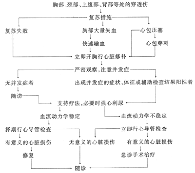
Figure 1 Diagnostic and treatment procedure for penetrating cardiac injury
Any patient with chest or abdominal trauma, where the estimated blood loss does not match the clinical condition, or who does not respond rapidly to adequate blood transfusion, should be highly suspected of having pericardial tamponade. Additionally, if initial stage [first stage] hypotension improves rapidly after blood volume replenishment but soon reappears, even leading to cardiac arrest, cardiac tamponade should be considered, and immediate surgical treatment is required.
When there is injury to the cardiac septum or heart valve membrane, corresponding heart murmurs can be heard in the precordial area or heart valve auscultation area, and even tremors can be palpated. Injury to the cardiac conduction system may result in bradycardia or conduction block.
Measurement of venous pressure is very helpful in distinguishing between pericardial tamponade and acute blood loss. An increase in central venous pressure is one of the early signs of pericardial tamponade. In cases of massive intrathoracic bleeding, before blood volume is corrected, the rise in venous pressure, jugular vein distension, and pulsus paradoxus may not be obvious, and even with complete circulatory failure, central venous pressure may remain normal. After rapid blood volume replenishment, central venous pressure may show an abnormal increase, and a value greater than 15cmH2O is diagnostically significant. Central venous pressure should be measured repeatedly. At the same time, the following should be ensured: ① the measurement zero point is properly adjusted; ② the water column in the tube should fluctuate with respiration during measurement; ③ the measurement should be taken when the patient is in a quiet state.
Echocardiography is very helpful in diagnosing pericardial tamponade, cardiac foreign bodies, hemopericardium, heart valve membrane, and ventricular septal perforation. It can also estimate the amount of pericardial effusion. However, when blood in the pericardium has coagulated, the misdiagnosis rate is higher.
Pericardiocentesis is very valuable for both the diagnosis and treatment of pericardial tamponade. However, when blood in the pericardial cavity has coagulated, false negatives may occur, which should be noted.
X-ray examination is not very helpful in diagnosing acute cardiac injury. However, chest X-rays can show hemothorax, pneumothorax, metallic foreign bodies, or injuries to other organs. If a fluid level is seen in the pericardium on a chest X-ray, it is diagnostically significant. Under fluoroscopy, heartbeats are weakened in cases of pericardial tamponade.
Electrocardiogram findings are generally atypical and not very helpful for diagnosis. If there is a decrease in voltage or changes in the S-T segment, it can assist in diagnosis.
For patients with diagnosed massive intrathoracic bleeding and suspected cardiac injury, emergency thoracotomy should be performed without the need for the above examinations, to avoid missing the opportunity for treatment.
bubble_chart Treatment Measures
Cardiac trauma should be primarily treated with surgery. Removing blood clots and accumulated blood from the pericardial cavity, and repairing and suturing the cardiac laceration can promptly relieve pericardial tamponade, control bleeding, and prevent complications such as pericarditis.
Treatment principle: Any penetrating cardiac injury with hemodynamic significance should be treated with surgery as soon as possible. Early relief of pericardial tamponade, control of bleeding, and prevention of complications are essential.
(1) Emergency Treatment
1. Anti-shock treatment: Place a central venous pressure catheter as soon as possible, rapidly transfuse blood and fluids intravenously to replenish blood volume and support circulation. This is a crucial step for successful emergency treatment. Vasopressors may be appropriately administered.
2. Maintain airway patency and support respiratory function: If the airway is not clear or the patient is unconscious, perform rapid tracheal intubation and artificial respiration. For patients with massive hemothorax or pneumothorax, perform closed thoracic drainage to promote lung expansion and improve breathing.
3. Pericardiocentesis: For confirmed pericardial tamponade, emergency pericardiocentesis should be performed, which can immediately improve the condition of some critically ill patients. However, if bleeding continues, the condition may still deteriorate. If the puncture needle is equipped with a plastic catheter, the catheter can be left in place until surgical decompression is performed to drain the accumulated blood from the pericardium.
During pericardiocentesis, the patient can be placed in a semi-recumbent position (30-50° tilt). The ideal puncture site is near the xiphoid process under the left costal margin. A fine pericardiocentesis needle or a plastic catheter-guided needle can be used. When using a metal needle, it can be performed under ECG monitoring by connecting the ECG electrode (chest lead) to the end of the puncture needle. When the needle touches the epicardium, an elevation of the S-T segment on the ECG can be observed, which helps avoid accidental injury to the myocardium during puncture. Pericardiocentesis can also be performed under ultrasound guidance. If no blood is aspirated during pericardiocentesis but the clinical diagnosis of pericardial tamponade is still highly likely, a subxiphoid pericardial window exploration should be rapidly performed under local anesthesia. Make a small midline incision below the xiphoid process, remove the xiphoid, push aside the pleural membranes, slightly incise the diaphragm, create a small window in the pericardium, insert a finger to explore the pericardial cavity, and then place a decompression drainage tube.
After emergency pericardiocentesis, surgical preparation should be made as soon as possible. Preoperative preparation mainly involves rapid and massive blood transfusion, supplemented by other anti-shock measures. If hypotension occurs, vasopressors (such as dopamine, isoproterenol, etc.) can be administered in appropriate amounts to increase myocardial contractility.
(2) Surgical Treatment
1. Surgical indications: Immediate surgery is required for myocardial penetrating injuries accompanied by pericardial tamponade or progressive hemorrhagic shock, or if pericardial tamponade signs reappear rapidly after pericardiocentesis. If circulation has stopped or the general condition is too poor, immediate thoracotomy should be performed in the emergency room. For other cases, after detailed examination, if there is a definite lesion, especially with symptoms of pericardial tamponade or bleeding causing a drop in blood pressure, surgical treatment is necessary.
2. Preoperative special management: If the penetrating object, such as a sharp knife, is still in the chest wall, it should not be hastily removed before surgery. If cardiac arrest occurs before surgery, emergency thoracotomy should be performed for cardiac compression to relieve pericardial tamponade, and the bleeding site should be temporarily controlled with a finger to improve cardiac output. External cardiac massage is not only ineffective but may also worsen pericardial tamponade.
3. Anesthesia: General anesthesia with tracheal intubation is preferred. At the start of surgery, administer a small amount of light anesthesia and provide ample oxygen. General anesthesia can dilate peripheral vessels, and positive pressure ventilation can further affect venous return, easily inducing cardiac arrest. Therefore, during anesthesia induction, be prepared for emergency thoracotomy, and avoid intermittent positive pressure ventilation before incising the pleural membrane. For critically ill, unconscious patients, anesthesia may be omitted or local anesthesia may be used.
4. Position and Incision: The patient is placed in a supine position with the injured side elevated by 30°. The skin of the anterior chest is widely disinfected. The choice of incision depends on the path and condition of the penetrating injury, and it must provide good exposure of the cardiac wound. The most commonly used incision is the left anterolateral thoracotomy, entering the chest through the fourth intercostal space. If necessary, the fourth and fifth costal cartilages can be severed to increase exposure. If the entry wound is on the right side, a right anterolateral incision is used. If exposure on one side is inadequate, the incision can be extended to the opposite side by transecting the sternum and ligating the internal thoracic vessels. If injury to major intrathoracic vessels is suspected, a median sternotomy is preferred. The aforementioned subxiphoid pericardial window, in addition to being used for diagnosis and emergency treatment, can also be performed initially when surgery is planned. If hemopericardium is found, the incision can be extended to perform a median sternotomy.
5. Cardiac Repair In cases of cardiac tamponade, the pericardial tension is extremely high. Once the pericardium is incised for decompression, blood gushes out, leading to immediate hemodynamic improvement in the patient. Rapid blood volume replenishment is essential. Enlarge the pericardial incision and remove blood clots. Expose the cardiac wound, temporarily stop the bleeding by pressing with a finger, and then proceed with repair suturing. Atrial wounds can often be controlled with atraumatic clamps. Large cardiac lacerations may cause further blood loss during suturing, necessitating rapid blood volume replenishment. Stabilize the circulation to allow ample time for wound repair.
There are many repair methods, and the choice depends on the specific situation.
(1) After pressing the laceration with a finger, use 1-0 or 2-0 Prolene sutures to pass through the full thickness of the myocardium at the fingertip area of the laceration, but do not penetrate the endocardial membrane (Figure 2). Slightly move the finger downward to expose the upper end of the laceration, and the assistant immediately ties the suture to approximate the edges of the laceration, achieving hemostasis without tearing the myocardium. Proceed with interrupted sutures step by step, gradually moving the pressing finger away until the entire laceration is closed.
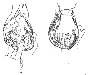
Figure 2: Cardiac Wound Suturing Method
A. Press the laceration with a finger, then pass the suture needle through both edges of the laceration for single-layer suturing;
B. The assistant stabilizes the heart with their hand, separating the fingers to expose the cardiac wound for the surgeon to suture and tie.
(2) After pressing the laceration with a finger, place traction sutures on both sides of the laceration (Figure 3). Cross and pull these traction sutures to achieve hemostasis, then perform interrupted suturing of the laceration under direct vision. The traction sutures can be removed or gently tied together.
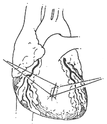
Figure 3: Cross-Traction Suture Hemostasis Method
Place parallel sutures on both sides of the laceration, then cross and pull the sutures to control bleeding and suture the laceration.
(3) For larger myocardial lacerations, temporarily stop the bleeding by plugging the laceration with a finger. First, place a purse-string suture around the laceration. Gradually withdraw the finger while gently tightening and tying the suture to close and reduce the laceration. Then, gently press the surface of the laceration to stop the bleeding. Continue suturing around the fingertip as described above, moving backward until the entire laceration is sutured.
If the laceration is near the coronary artery, use a pericardial or Dacron patch as a bolster for mattress sutures. Pass the suture needle under the coronary artery through the myocardium to suture the laceration, avoiding injury to the coronary artery (Figure 4).
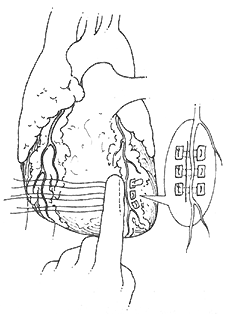
Figure 4: Mattress Suture for Cardiac Laceration
When the laceration is adjacent to a coronary vessel, use mattress sutures, passing the suture needle under the finger and coronary vessel through the myocardium to suture the laceration (the figure shows the completed suturing).
(4) If the myocardial laceration is too large to be directly sutured, use pericardium or pedicled muscle to fill the defect, followed by mattress suturing. Alternatively, occlude the superior and inferior vena cava for 60-90 seconds to reduce cardiac blood volume, quickly place sutures, and tie them during the next occlusion. The most reliable method is to immediately establish cardiopulmonary bypass for repair. In cases with a left thoracotomy, insert a venous return cannula into the right ventricular outflow tract or pulmonary artery, and insert an arterial supply cannula into the descending aorta or femoral artery. Repair the myocardial laceration with a Dacron patch.
(5) Directly repair coronary artery lacerations with 6-0 Prolene sutures. If ligation is required due to rupture, and distal blood supply is compromised, leading to myocardial pallor, perform a coronary artery bypass graft using the saphenous vein or internal thoracic artery.
(6) If a wound is suspected on the posterior wall of the heart, the pericardium should be widely incised, and the heart should be gently lifted with the hand to expose the posterior wall and locate the tear for repair. Under conditions of hypoxia, acidemia, and hypovolemia, moving the heart can easily cause arrhythmias and cardiac arrest, so special attention should be paid. Due to technical reasons, wounds on the posterior wall of the atrium are not easily sutured directly and can be managed with a tamponade hemostasis method.
(7) The atrial tear is clamped with a non-traumatic vascular clamp and then sutured. Generally, 4-0 or 3-0 silk thread is used for interrupted or continuous suturing.
(8) If cardiac arrest occurs when opening the pericardium, immediate cardiac massage should be performed, and 2-3 ml of 1:1000 adrenaline should be injected into the cardiac chamber. At the same time, the left lung should be retracted forward to expose the descending aorta, and the descending aorta should be clamped to facilitate blood supply to the coronary arteries and brain. Repair the wound as quickly as possible. When the heart resumes strong beating, gradually release the clamp on the descending aorta, taking care not to injure the esophagus.
(9) Most injuries to the vena cava can be clamped and sutured tangentially for hemostasis. If direct suturing is not possible, an internal shunt can be performed through the right atrial appendage before suturing and repairing (Figure 5).
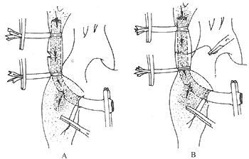
Figure 5: Internal shunt suturing method for vena cava injury
A side-hole plastic tube is inserted through the right atrial appendage into the distal side of the tear for internal shunting and bleeding control; B. Suturing the superior vena cava tear
(10) Repair of intracardiac structures: After repairing the cardiac wall wound, routinely palpate the cardiac wall. The presence of a tremor is a sign of intracardiac structural injury, which may be due to perforation of the atrial or ventricular septum or damage to the heart valve. Unless the damage to the intracardiac structures directly affects the patient's survival and requires immediate repair, it is generally not repaired immediately. After the patient's condition stabilizes postoperatively, the diagnosis should be confirmed by echocardiography, cardiac catheterization, or angiography before repair. Intracardiac structural damage without hemodynamic significance may not require repair. Whether for emergency repair or secondary surgical repair, it should generally be performed under cardiopulmonary bypass with direct vision.
(11) Removal of cardiac foreign bodies: The success of removing cardiac foreign bodies depends not only on accurate localization but also on the surgical technique. Since the heart is a moving organ, metallic foreign bodies such as bullets can remain in the myocardium or cardiac chambers and may shift with blood flow, posing a risk of pulmonary or systemic embolism. During surgery, the position of the foreign body may change due to manipulation. Depending on the type, size, and location of the foreign body, different methods should be used for removal. For example, clamping the exposed part of the foreign body outside the heart and removing it; temporarily blocking the blood supply to the myocardium, incising the heart wall to remove it; using a finger to press the foreign body inside the cardiac chamber, incising the heart wall, and clamping to remove the foreign body. Or removing the foreign body under cardiopulmonary bypass. In summary, when removing cardiac foreign bodies, be vigilant about the shifting of the foreign body, and avoid excessive manipulation. Random palpation may shift the foreign body, increasing the difficulty of removal.
When repairing the wound, carefully check for any missed fistula disease wounds and explore for any atrial septal injury. Thoroughly clean the pericardial cavity, loosely suture the pericardium, and create a window for drainage to prevent recurrent pericardial tamponade. Postoperatively, routinely administer tetanus antiserum and antibiotics to prevent infection, and closely monitor blood pressure, heart rate, and central venous pressure. Tonify blood, replenish fluids, and expand volume. Follow-up should also be conducted postoperatively to prevent complications such as traumatic ventricular aneurysm, coronary artery fistula, or coronary artery aneurysm, as well as constrictive pericarditis.
Surgical outcomes
Since Rehn first successfully sutured a cardiac stab wound in 1896, there has been significant controversy over the treatment methods for penetrating cardiac injuries. Blalock and others favored pericardial puncture treatment, while other scholars advocated for surgical treatment or prompt surgical intervention after failed pericardial puncture. It was only in recent years that the view that penetrating cardiac injuries should primarily be treated surgically has been widely recognized by scholars worldwide. In recent years, the reported surgical outcomes for penetrating cardiac injuries vary, with mortality rates ranging from 5% to 20%. The mortality rate mainly depends on the type of injury, the patient's circulatory status upon arrival at the hospital, and whether cardiac arrest occurs during thoracotomy.
Most (60-80%) patients with open cardiac injuries die shortly after the injury due to acute cardiac tamponade and massive hemorrhage. Therefore, timely and effective emergency measures are crucial for the patient's survival. Small injuries to the pericardium and myocardium can heal on their own. For example, small lacerations (<0.5~1cm) of the pericardium and myocardium caused by sharp objects such as knives often result in self-limiting intrapericardial bleeding or can be cured after pericardial puncture and decompression. In contrast, gunshot wounds to the pericardium or large cardiac wounds cause rapid and severe bleeding, requiring immediate surgical repair and suturing, but patients often do not survive long enough to reach the hospital.
Injuries involving two cardiac chambers are more severe than those involving a single chamber. Among 228 cases of comprehensive surgery, the overall mortality rate reached 79%, with a cure rate of only 21%. Among the fatal cases, simultaneous injuries to the left atrium and left ventricle were the highest (93%), while injuries to the right atrium and left atrium were lower (55%). Combined injuries with other organs further exacerbate the condition and increase mortality. Among the associated injuries, lung injuries were the most common, followed by the liver, esophagus, spleen, stomach, and inferior vena cava.
From 1984 to 1993, Beijing Anzhen Hospital admitted a total of 9 patients with penetrating cardiac injuries, of which only 1 died due to prolonged hypotension and postoperative cerebral complications, while the remaining 8 were cured. Two typical cases are briefly described below:
One case involved a stab wound to the chest wall with four wounds. The patient was admitted as an emergency and underwent surgery. Upon left anterolateral thoracotomy, a 2.5 cm wound was found above the apex of the right ventricle, and a 3.0 cm wound at the apex of the left ventricle, with continuous bleeding. The surgery involved clearing the pericardial blood, using gauze to press the bleeding at the left ventricular apex, and first suturing the right ventricular wound with 4-0 Prolene continuous sutures. The left ventricle was gently lifted, the wound pressed with the thumb, and three interrupted mattress sutures with 3-0 Prolene were applied to stop the bleeding. Postoperatively, blood transfusion was accelerated, blood pressure stabilized, and the patient recovered.
Another case also involved a stab wound, entering from the third intercostal space at the left sternal border. Upon admission, blood pressure was stable, chest X-ray showed left thoracic hemothorax, no blood in the pericardial cavity, and no heart murmurs on auscultation. Two thoracenteses were performed on the left chest, extracting 400ml and 200ml of blood respectively. Twelve hours post-injury, a significant systolic heart murmur and systolic tremor appeared, and echocardiography confirmed a ventricular septal rupture with left-to-right shunt. Fourteen days post-injury, a ventricular septal rupture repair was performed under general anesthesia and extracorporeal circulation. No pericardial blood was found during surgery, the wound was located at the lower part of the right ventricular outflow tract without bleeding, and the right ventricular outflow tract was incised to reveal thickened ventricular septal muscle. The stab wound was at a 30° angle to the septum, and interrupted mattress sutures with 5×14 silk and continuous sutures with 4-0 Prolene were applied. The patient recovered smoothly.
Therefore, any penetrating injury within the dangerous zone of the heart, whether acute or mild, should be promptly diagnosed, managed, and closely monitored. Once the condition changes, surgery is necessary to save the patient's life.


