| disease | Inguinal Oblique Hernia |
| alias | Inguinal Hernia |
When internal organs or tissues protrude through the inguinal canal, it is called an indirect inguinal hernia, accounting for about 90% of inguinal hernias and being the most common type of external hernia.
bubble_chart Etiology
During embryonic development, the fetal testes are located below the kidneys in the abdominal {|###|} membrane, with their lower ends connected to the scrotum by the gubernaculum. As the fetus grows, the testes gradually descend through the inguinal canal into the scrotum. The abdominal {|###|} membrane, closely attached to the front of the testes, also extends downward to form a sac-like structure, enveloping the testes and forming the processus vaginalis of the abdominal {|###|} membrane. Before birth, the portion of the processus vaginalis closely attached to the testes becomes the tunica vaginalis of the testes, while the remainder closes off and atrophies into a fibrous cord. If the processus vaginalis fails to close and remains connected to the abdominal cavity, it becomes a congenital hernial sac, allowing abdominal organs or tissues to protrude through the residual processus vaginalis and the inguinal canal, forming an indirect inguinal hernia. In females, the round ligament of the uterus passes through the inguinal canal, leading to a similar protrusion of the abdominal {|###|} membrane that descends into the labia majora. If this fails to close, it can also result in an indirect inguinal hernia.
Additionally, congenital developmental defects that impair the physiological closure mechanism of the inguinal canal are another significant disease cause of indirect inguinal hernias.
bubble_chart Clinical Manifestations
The presence of a reducible mass at the external inguinal ring is the most important clinical manifestation. Initially, the mass protrudes obliquely along the inguinal canal toward the external ring during prolonged standing, walking, or coughing. Over time, the mass gradually enlarges and extends into the scrotum. The upper part of the mass is narrow, while the lower part is broad, giving it a pear-like shape, with a stalk-like extension into the inguinal canal. When the mass protrudes, there is a sensation of heaviness or grade I dull pain.During examination, the patient should lie supine with the affected hip flexed and adducted to relax the inguinal region. Gently pressing the mass upward and outward along the inguinal canal can reduce it. If pressure is applied 2 cm above the midpoint of the inguinal ligament to compress the internal ring and the patient is asked to stand and cough, the protrusion of the mass can be prevented. Removing the pressure allows the mass to reappear. In cases of incomplete indirect hernia, where the hernia contents have not protruded through the external ring, a finger can be inserted into the external ring. Asking the patient to cough will elicit an impulse sensation.
In cases of irreducible hernia, the mass is difficult or only partially reducible during examination.
If the mass cannot be reduced after protrusion and becomes incarcerated, the protruding hernia will cause severe pain, high tension, and tenderness. If the hernia contents include intestine, clinical manifestations of acute mechanical intestinal obstruction will occur.
If incarceration is not relieved, the blood supply to the hernia contents becomes compromised, leading to strangulated hernia. Intestinal ischemia and necrosis will occur, and the hernia mass will exhibit signs of acute inflammation such as redness, swelling, heat, and tenderness, along with signs of peritonitis. In some cases, systemic infection, high fever, fear of cold, and other symptoms may become very pronounced, and severe cases may develop septic shock.
bubble_chart Treatment MeasuresAn indirect inguinal hernia cannot heal on its own and may become incarcerated or strangulated, thus requiring surgical treatment. However, for infants under one year old, the abdominal wall may strengthen with growth and development, potentially allowing spontaneous healing, so surgery can be temporarily postponed. Elderly or frail patients with other severe conditions unsuitable for surgery may use a truss to compress the hernia ring after reducing the hernia, removing it during nighttime rest. Long-term use of a truss can cause adhesions between the hernia contents and the neck of the hernia sac, which is generally not recommended.
The surgical principle for indirect inguinal hernia involves high ligation of the hernia sac and hernia repair. For infants, only high ligation of the hernia sac is performed to avoid affecting the development of the spermatic cord and testis or disrupting the physiological closure mechanism of the inguinal canal. Hernioplasty is rarely performed unless there is a significant defect in the abdominal wall.
High ligation of the hernia sac: To eliminate the residual processus vaginalis of the peritoneum, after transecting the hernia sac, its proximal end must be dissected to the internal ring, where the extraperitoneal fat layer can be seen, with the parietal peritoneum lying beneath. At this level, the neck of the hernia sac is ligated with silk sutures, and the distal sac is usually left open without resection.
Hernia repair: As an indirect inguinal hernia progresses, the internal ring gradually enlarges, further weakening the abdominal wall. Therefore, after high ligation of the hernia sac, hernia repair must be performed. Hernia repair should include two concepts: repairing the enlarged internal ring and reinforcing the weak areas of the inguinal canal. Before repairing the inguinal canal, the enlarged internal ring must first be explored and repaired; otherwise, recurrence is inevitable. To achieve this, after high ligation of the hernia sac, the cremaster muscle must be further dissected and transected at its root to better expose the enlarged internal ring and the interfoveolar ligament. The interfoveolar ligament is sutured to reduce the internal ring to a size that only allows the spermatic cord to pass through. The main techniques for repairing the weak areas of the inguinal canal include the following:
1. Ferguson method (Figure 1): The lower edge of the internal oblique muscle, the transversus abdominis aponeurotic arch, and the conjoined tendon are sutured superficial to the spermatic cord to the inguinal ligament to reinforce the anterior wall of the inguinal canal. This method is suitable for smaller hernias with an intact posterior wall of the inguinal canal.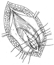
(1) The spermatic cord remains in its original position, and the cremaster muscle is sutured.
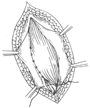
(2) The lower edge of the internal oblique muscle, the transversus abdominis aponeurotic arch, and the conjoined tendon are sutured to the inguinal ligament.
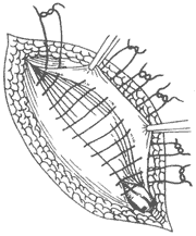
(3) The external oblique aponeurosis is overlapped and sutured.
Figure 1 Ferguson procedure
2. Bassini method (Figure 2): The spermatic cord is freed and lifted, and the lower edge of the internal oblique muscle, the transversus abdominis aponeurotic arch, and the conjoined tendon are sutured deep to the spermatic cord to the inguinal ligament to reinforce the posterior wall of the inguinal canal. The spermatic cord is repositioned between the internal oblique muscle and the external oblique aponeurosis. This method is suitable for larger hernias with a weakened posterior wall of the inguinal canal. The strength of the posterior wall of the inguinal canal, the transversus abdominis aponeurosis, and the transversalis fascia can be assessed intraoperatively by inserting a finger into the internal ring and pressing the abdominal wall toward the body surface to evaluate its strength. This technique is currently more commonly used.
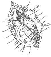
(1) The spermatic cord is lifted and repositioned between the internal oblique muscle and the external oblique muscle. Deep to it, the lower edge of the internal oblique muscle, the transversus abdominis aponeurotic arch, and the conjoined tendon are sutured to the inguinal ligament.
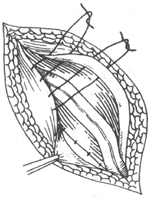
(2) The external oblique aponeurosis is overlapped and sutured.
Figure 2 Bassini procedure
3. Halsted's Method (Figure 3) The spermatic cord is freed and lifted, and beneath it, the lower edge of the internal oblique muscle, the aponeurotic arch of the transversus abdominis, and the conjoined tendon are sutured to the inguinal ligament. Then, the upper and lower leaves of the external oblique aponeurosis are approximated or overlapped and sutured beneath the spermatic cord, which is displaced subcutaneously. This technique further reinforces the posterior wall of the inguinal canal compared to the Bassini method. The indications are the same as those for the Bassini method, but it is generally not suitable for adolescents, as subcutaneous displacement of the spermatic cord may affect the development of the cord and testes.
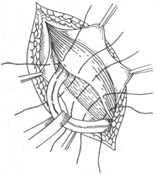
(1) Lift the spermatic cord, shift it subcutaneously, and suture the lower edge of the internal oblique muscle, the aponeurotic arch of the transversus abdominis, and the conjoined tendon to the inguinal ligament.
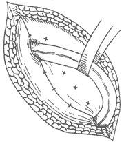
(2) Overlap and suture the external oblique aponeurosis deep to the spermatic cord.
Figure 3 Halsted's operation
4. McVay method (Figure 4) The pectineal ligament (Cooper's ligament) is used instead of the inguinal ligament in the Bassini method for repair. The transversalis fascia is incised along the posterior wall of the inguinal canal and the upper edge of the inguinal ligament. The upper cut edge, along with the lower edge of the internal oblique muscle, the aponeurotic arch of the transversus abdominis, and the conjoined tendon, is sutured to the pectineal ligament to restore the original normal anatomical relationship. The repair site extends deep to the superior pubic ramus, not only reinforcing the posterior wall of the inguinal canal but also altering the direction of intra-abdominal pressure transmission. This method is suitable for large indirect and direct hernias. However, it should be noted that this procedure does not simultaneously close the internal ring. If the internal ring is significantly enlarged, it should still be repaired, or the upper cut edge of the transversalis fascia should be sutured to the anterior wall of the femoral sheath to narrow the internal ring to a size just allowing the passage of the spermatic cord. This repair is deep and technically challenging, and care must be taken to avoid injury to the femoral vessels.
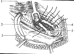
Figure 4 McVay's operation
1. Internal oblique muscle 2. Inguinal ligament 3. External oblique aponeurosis 4. Aponeurotic arch of the transversus abdominis 5. Pectineal ligament
The figure shows suturing the lower edge of the internal oblique muscle and the aponeurotic arch of the transversus abdominis to the pectineal ligament, followed by suturing the transversalis fascia to reconstruct the internal ring to a size just allowing the passage of the spermatic cord.
5. Preperitoneal repair The advantage of this procedure is that the hernia sac can be ligated at a higher position without disrupting the anatomical structure of the inguinal canal or its physiological closure mechanism. It does not require incision of the transversalis fascia in the inguinal canal area to suture the lower edge of the internal oblique muscle, the aponeurotic arch of the transversus abdominis, and the conjoined tendon to the inguinal ligament or pectineal ligament. It is particularly suitable for recurrent inguinal hernias, as it avoids adhesions and scar tissue from previous surgeries. Specific steps: Use the Nyhus approach to make a transverse incision about 6 cm above the inguinal canal through the external oblique aponeurosis, internal oblique muscle, transversus abdominis muscle, and transversalis fascia. Dissect downward deep to the preperitoneal fascia to locate the neck of the hernia sac, incise the sac wall, reduce the hernia contents, perform high ligation of the hernia sac, and carry out preperitoneal hernia repair. For recurrent inguinal hernias with severe defects in the inguinal region, autologous fascia lata or synthetic mesh can be used for repair. The medial lower edge of the graft is sutured to the pectineal ligament, crossing the femoral vessels and continuing laterally to the inguinal ligament and iliopubic tract. The lateral edge of the mesh is cut into a bifurcated shape to encircle the spermatic cord and reconstruct the internal ring. The upper and medial edges of the mesh are sutured to the transversalis fascia, transversus abdominis muscle, and rectus abdominis muscle, respectively.
6. Shouldice Method (Figure 5) The principle involves excising the weakened transversalis fascia, overlapping its upper and lower leaves in a imbricated manner, suturing the edge of the upper leaf to the inguinal ligament, and then suturing the conjoined tendon, the transversus abdominis aponeurotic arch, the lower border of the internal oblique muscle, and the deep surface of the lower leaf of the external oblique aponeurosis to the inguinal ligament. The specific procedure involves mobilizing and elevating the spermatic cord, inserting a finger into the internal ring to assess the extent and severity of transversalis fascia weakness, and incising the transversalis fascia along the inguinal ligament from the internal ring to the pubic tubercle. The weakened portion is excised, and the lower leaf is freed to the inguinal ligament, while the upper leaf is freed medially to the posterior rectus sheath. The intact upper and lower leaves are then imbricated, with the cut edge of the lower leaf continuously sutured from the pubic tubercle outward to the deep surface of the upper leaf until a snug internal ring is formed, just allowing passage of the spermatic cord. The suture is then reversed in an antagonistic direction to secure the cut edge of the upper leaf to the inguinal ligament and tied back to the pubic tubercle with the other end of the initial suture. Next, the lower border of the internal oblique muscle, the transversus abdominis aponeurotic arch, and the conjoined tendon are sutured to the inguinal ligament and the deep surface of the external oblique aponeurosis. Finally, the external oblique aponeurosis is sutured superficial to the spermatic cord. This method emphasizes reinforcing the role of the transversalis fascia in hernia repair and is suitable for indirect hernias with weakened posterior inguinal wall, thin transversalis fascia, and an enlarged internal ring.
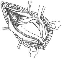 | 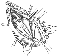 |
| (1) Lift the spermatic cord and incise the transversalis fascia along the dotted line. | (2) Free the upper and lower flaps of the incised edge of the transversalis fascia. |
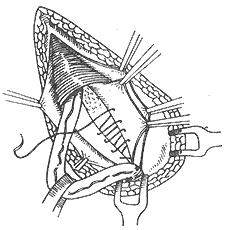 | 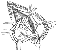 |
| (3) Continuously suture the lower incised edge of the transversalis fascia from the pubic tubercle upward and outward to the deep surface of the upper flap. | (4) Then, in the antagonistic direction, continuously suture the upper incised edge of the transversalis fascia to the inguinal ligament until reaching the pubic tubercle. |
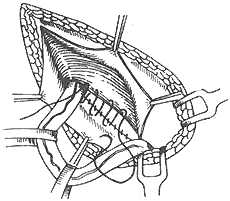 | 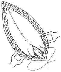 |
| (5) Suture the lower edge of the internal oblique muscle, the aponeurotic arch of the transversus abdominis, and the conjoined tendon to the inguinal ligament or the deep surface of the external oblique aponeurosis. | (6) Suture the external oblique aponeurosis superficial to the spermatic cord. |
Figure 5 Shouldice Procedure
7. Madden Method (Figure 6) This procedure only repairs the transversalis fascia. After freeing and lifting the spermatic cord, insert a finger into the internal ring to assess its size, the degree of weakness in the transversalis fascia, and the extent of the defect. Incise the transversalis fascia along the inguinal ligament from the internal ring, dissect the upper and lower flaps of the transversalis fascia to healthy tissue, excise the weakened portion, and then intermittently suture the edges of the two flaps from the lacunar ligament outward to the root of the spermatic cord, reconstructing the internal ring. The procedure is similar to the Shouldice method, emphasizing the importance of reinforcing the transversalis fascia but without repairing other layers of the abdominal wall, which aligns more closely with anatomical principles. Since the sutured area of the transversalis fascia has minimal tension, patients experience no pulling sensation postoperatively. However, this technique is not suitable for large indirect hernias where the transversalis fascia and the strength of the inguinal region are severely compromised.
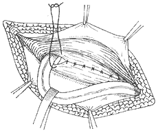
Figure 6 Madden Procedure
Lift the spermatic cord, incise the transversalis fascia along the inguinal ligament from the internal ring, and excise its weakened portion. Then, intermittently suture the edges of the upper and lower flaps from the lacunar ligament outward to the root of the spermatic cord, reconstructing the internal ring.
Hernioplasty: For large indirect hernias with severe weakness and defects in the posterior wall of the inguinal canal, where the transversus abdominis aponeurotic arch, transversus abdominis muscle, and internal oblique muscle have atrophied and cannot be utilized for repair, autologous fascia lata, silk mesh, or various synthetic fiber meshes can be used for hernioplasty. Alternatively, the anterior sheath of the rectus abdominis can be turned outward and downward and sutured to the inguinal ligament to reinforce the posterior wall of the inguinal canal.
Incarcerated hernia requires emergency surgery, which involves incising the constricted hernial ring, relieving the incarceration, reducing the hernial contents, and performing high ligation of the hernial sac. If there is no edema in the local tissues, herniorrhaphy can be performed simultaneously.
For pediatric incarcerated indirect inguinal hernia, non-surgical treatment may be attempted first. However, if it is a strangulated indirect inguinal hernia, emergency surgery is required regardless of age. The goals of surgery are to relieve the incarceration, resect the necrotic hernial contents, and perform high ligation of the hernial sac. Herniorrhaphy is contraindicated. Preoperative preparation is crucial to enhance the safety of surgery for strangulated indirect inguinal hernia.
Combined with the anatomical characteristics of the inguinal region and the aforementioned clinical manifestations, the diagnosis of indirect inguinal hernia is not difficult. However, it must be differentiated from the following diseases:
- Hydrocele of the tunica vaginalis: A positive transillumination test of the mass is a characteristic clinical manifestation of this condition. Additionally, the mass has a clear boundary, and its upper pole does not connect to the external ring. If the testis is encased in the hydrocele, it may be difficult to palpate. The mass cannot be reduced, and there is no history of reducibility. If the processus vaginalis peritonei is not completely closed, forming a communicating hydrocele of the tunica vaginalis, although the mass may also exhibit reducibility, it can be differentiated using the transillumination test.
- Cyst of the round ligament of the uterus: The mass is located in the inguinal canal, round or oval in shape, with a cystic sensation, clear boundaries, and high tension. Its upper end does not extend into the abdominal cavity, and it is generally not easily confused with a hernia.
- Cyst of the spermatic cord or incomplete descent of the testis: The mass is located along the inguinal canal or the path of the spermatic cord and testis, with clear boundaries. The former has a cystic sensation and high tension, and the ipsilateral testis can be palpated in the scrotum. The latter is firm and solid in texture, with the absence of the ipsilateral testis in the scrotum.


