| disease | Internal Hernia |
Internal hernias occur when abdominal organs or omenta shift from their original positions through normal or abnormal openings or fissures within the abdominal cavity.
bubble_chart Etiology
During embryonic development, after the midgut rotates 270° counterclockwise, the cecum becomes fixed in the right iliac fossa. The root of the mesentery of the midgut fuses with the posterior abdominal wall, forming peritoneal folds or recesses near the duodenum, cecum, and the root of the sigmoid mesocolon. If these recesses are large and deep, or if the opening (Winslow’s foramen) left during the formation of the omental bursa (lesser peritoneal cavity) is wide, intestinal loops may herniate through them. After the 10th week of embryonic development, when the midgut returns to the abdominal cavity, the small intestine may also herniate into the mesentery of the midgut loop, forming an internal hernia. Additionally, acquired factors such as postoperative adhesive bands or abnormal spaces created by gastrointestinal anastomoses can allow intestinal loops to herniate.
Internal hernias can be classified into true and false hernias based on the presence or absence of a hernial sac. When organs pass through normal or abnormal openings into another peritoneal sac or omental bursa, it is called a true hernia due to the presence of a sac. If the omentum or mesentery develops abnormal fissures during embryonic development, or if an abnormal opening is created due to abdominal surgery, allowing intestinal loops to herniate without a sac, it is called a false hernia.
**Paraduodenal Hernia** One of the most common congenital internal hernias (Figure 1). Left-sided hernias are more frequent, where intestinal loops herniate into the left paraduodenal recess (Landzert’s recess) near the ascending duodenum. The opening faces right, bounded superiorly by the duodenojejunal flexure, the inferior border of the pancreas, and the origin of the left renal vessels. The anterior boundary is formed by the inferior mesenteric vein and the left colic artery, while the right boundary is the main trunk of the superior mesenteric artery. The hernial sac extends leftward, with the descending mesocolon superficially and the left kidney, ureter, and psoas major deeply. Right-sided paraduodenal hernias involve intestinal loops herniating into the recess below the horizontal duodenum and duodenojejunal flexure (Waldeyer’s recess). This recess opens leftward, bounded superiorly by the duodenum, posteriorly by the lumbar vertebrae, and anteriorly by the superior mesenteric vessels. The hernial sac extends rightward, with the ascending and transverse mesocolon superficially and the right kidney, ureter, inferior vena cava, and psoas major deeply.
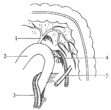
(1) Hernial orifice
1. Middle colic artery 2. Duodenum 3. Superior mesenteric artery and vein 4. Inferior mesenteric vein 5. Left colic artery
① Landzert’s recess on the left side of the ascending duodenum
② Waldeyer’s recess below the horizontal duodenum
(2) Right paraduodenal hernia herniating from Waldeyer’s recess
**Figure 1 Paraduodenal hernia**
**Paracecal Hernia** Much rarer than paraduodenal hernias. Intestinal loops may herniate through the following recesses: the ileocolic recess above the ileum on the medial side of the ascending colon; the ileocecal recess below the ileocecal junction; and the cecal recess posterior and inferior to the cecum. The openings of these recesses contain branches of the ileal vessels and the herniated intestinal loops. The hernial sac lies in the space behind the cecum and ileocecal region.
**Sigmoid Mesocolon Hernia** Extremely rare. Intestinal loops herniate through the recess between the root of the sigmoid mesocolon and the posterior abdominal wall. The anterior border is formed by the sigmoid vessels, and the hernial sac extends inferolaterally in a funnel shape, with the sigmoid mesocolon superficially and the common iliac vessels and ureter deeply.
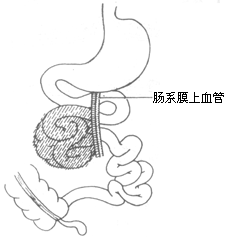
**Hernia of Winslow’s Foramen** Also rare. Intestinal loops herniate through Winslow’s foramen, with the omental bursa serving as the hernial sac (Figure 2).
**Figure 2 Hernia of Winslow’s foramen**
Small intestine herniating through Winslow’s foramen, with the omental bursa as the hernial sac.
Other internal hernias. Congenital intra-abdominal pseudohernias refer to internal hernias where the intestinal tract herniates through defects in the greater omentum, small intestine, or mesocolon, with herniation through defects in the mesentery of the small intestine being the most common. The most frequent site of occurrence is near the mesentery of the terminal ileum. Acquired intra-abdominal hernias are all pseudohernias and can be seen in the following scenarios: posterior to the anastomosis site in Billroth II subtotal gastrectomy with gastrojejunostomy; between a colostomy and the lateral abdominal wall; or within adhesive bands formed between loops of intestine or between the intestine and the abdominal wall, where the intestine herniates through these acquired gaps.
bubble_chart Clinical ManifestationsIf the diameter of the recess entrance is large, allowing the intestine to move freely in and out, some congenital internal hernias may be asymptomatic. However, chronic clinical manifestations of intestinal obstruction, such as {|###|}abdominal distension and fullness{|###|}, {|###|}nausea{|###|}, and {|###|}dull pain{|###|}, are usually present. When the pain is severe, a cystic mass may be palpable, which produces a tympanic sound upon percussion. During the {|###|}stage of remission{|###|}, a barium meal examination may reveal a cluster of {|###|}small intestine{|###|} coiled and fixed in one location. In cases of acute obstruction, a plain abdominal X-ray shows a cluster of {|###|}small intestine{|###|} fixed in one position with multiple fluid levels. If strangulation occurs, symptoms of strangulated intestinal obstruction and signs of peritonitis appear.
Congenital internal hernias are rare and lack distinctive clinical manifestations, making diagnosis difficult. They are often discovered during surgery for acute {|###|}small intestine{|###|} obstruction. Additionally, if severe acute {|###|}small intestine{|###|} obstruction occurs after abdominal surgery, the possibility of an acquired internal hernia should be considered.
bubble_chart Treatment Measures
All intra-abdominal hernias require surgical treatment. In congenital intra-abdominal hernias, the hernia ring margin often contains important blood vessels or organs. When reducing the intestine, forceful dilation or arbitrary cutting of the hernia ring should be avoided to prevent injury. For Winslow's foramen hernias, a Kocher incision can be made to fully mobilize the duodenum and enlarge the hernia ring. Paraduodenal hernias can only be incised below the hernia ring, especially for right-sided paraduodenal hernias, where care must be taken not to accidentally injure the mesenteric vessels on the anterior edge of the hernia ring. In summary, the anatomical relationships adjacent to the hernia ring must be carefully noted during surgery.
True congenital intra-abdominal hernias have a hernia sac composed of vascular-rich peritoneum, omentum, or mesentery, which can only be incised in avascular areas or away from major vessels to reduce and examine the incarcerated intestine. If the intestine is not necrotic but difficult to reduce due to distension, intestinal decompression can be performed under strict aseptic conditions before reduction. If strangulation and necrosis have occurred, the intestine should be transected at the normal segment near the hernia ring entrance, the necrotic segment removed from the hernia sac, and the ends anastomosed. The surgical principle for congenital or acquired intra-abdominal pseudoceles is to remove adhesions or suture all fissures after reducing the hernia contents to prevent recurrence.


