| disease | Congenital High Scapula |
| alias | Sprengel's Deformity |
This disease is a relatively rare congenital malformation. Its characteristics include an abnormally high position of the scapula, with the affected shoulder joint higher than the healthy side, limited upward movement of the affected arm, and possible concurrent deformities of the ribs, neck, or thoracic vertebrae. It was first described by Enlenber in 1863. In 1891, Sprengel reported four cases and discussed the disease's etiology, hence the condition is also known as Sprengel's deformity.
bubble_chart Etiology
This is the result of incomplete descent of the scapular leukorrheal disease during the embryonic period. The shoulder girdle is a limb bud adjacent to the cervical vertebrae during the embryonic period, gradually descending from the corresponding positions of C4 to C6 to the 2nd to 7th intercostal spaces starting from the fourth month of the embryo. Due to certain reasons, if the normal descent process of the shoulder girdle is obstructed, it leads to the formation of a high scapular deformity. It can occur unilaterally or bilaterally.
bubble_chart Pathological Changes
During embryonic development, the shoulder girdle descends, and the ratio of the transverse to vertical diameter of the scapula gradually decreases. However, due to interruption or obstruction of the descent process, the scapula remains in a higher position posterior to the thorax, affecting its normal development and leading to morphological changes. Common pathological changes can be divided into two aspects: ① Changes in bones and muscles. The former involves the scapula being positioned high, sometimes even contacting the occipital bone at its highest point. The upper part curves forward beyond the top of the thorax, forming a hook-like shape, while the inner edge and lower angle shift inward toward the spine, sometimes forming bony, cartilaginous, or fibrous connections with adjacent cervical or upper thoracic spinous processes. A complete bony connection is termed an omo-vertebral bone. Between the superomedial angle of the scapula and the cervical spinous or transverse processes, there may be a fibrous band or cartilaginous/bony bridge, known as the omo-vertebral bridge. In some cases, a well-developed joint exists between the bridge and the scapula, while in others, only fibrous tissue connects them. The scapular body is generally underdeveloped. Besides scapular deformities, associated anomalies may include scoliosis, vertebral body absence, rib fusion, and narrowed intercostal spaces. ② Muscular changes. Some or all of the scapular muscle groups may be absent, with the levator scapulae and rhomboid muscles becoming thin and exhibiting varying degrees of contracture or fibrosis.
bubble_chart Clinical ManifestationsThe clinical manifestations primarily include the observation of an elevated affected shoulder in children after the age of 1. "Elevated" refers to the relative position of the scapula and the rib cage, presenting as a shrugged-shoulder and short-neck appearance. There is significant limitation in shoulder joint abduction and elevation, underdeveloped shoulder girdle muscles on the affected side, and older patients may exhibit combined spinal and thoracic deformities. The scapula is underdeveloped, with an elevated lower angle, shortened vertical diameter, and widened transverse diameter. The limitation in shoulder joint abduction and elevation is closely related to the position and developmental deformity of the scapula, such as: ① The height of the shoulder girdle exceeds that of the rib cage, with the superior medial angle even bending forward. ② The medial border of the scapula is tightly adjacent to the vertebral spinous processes. ③ Cleido-vertebral bone. ④ Abnormalities in the muscles surrounding the scapula. X-ray findings may reveal a smaller and underdeveloped scapula on the affected side, an elevated lower angle, an upper border that may exceed the height of the rib cage, an increased width (transverse diameter) between the axillary and vertebral borders of the scapula, the lower angle turning toward the axilla, and the superior medial border turning toward the spine. Bone bridges connecting the scapula to the spine, as well as other cervicothoracic vertebral and rib deformities, may also be observed.
Functional impairment depends on the severity of the deformity. Cavendish classified the deformity into four grades based on severity: - **Grade I**: The deformity is not obvious, with both shoulders at the same level, and the appearance is nearly normal when clothed. - **Grade II**: Mild deformity, with both shoulders nearly level, but the deformity is visible when clothed, and a raised mass may be seen at the neck web. - **Grade III**: Moderate deformity, with the affected shoulder joint 2–5 cm higher than the contralateral side, making the deformity easily noticeable. - **Grade IV**: Severe deformity, with the affected shoulder significantly elevated, and the superior medial angle of the scapula nearly touching the occipital bone. Short-neck deformity may also be present. The grading of the deformity provides important reference for treatment.
bubble_chart Treatment MeasuresFor cases where the deformity is not severe and functional impairment is not significant, surgical treatment is not considered. Passive and active upper limb movements, such as abduction, elevation, depression, and adduction, can be performed to stretch and traction the shortened muscles, thereby improving and enhancing shoulder abduction and elevation functions.
Surgical treatment is suitable for children with severe deformity and obvious functional impairment. In addition to the elevated scapula, these patients often have other bony and soft tissue deformities. Therefore, the following factors should be considered when opting for surgical treatment: ① Age: The optimal surgical outcomes are typically achieved between the ages of 3 and 7. Surgery is not well-tolerated in younger children. For those over 8 years old, excessive focus on correcting the deformity during surgery often leads to brachial plexus nerve traction and injury. Additionally, as tissues are nearing maturity and lack elasticity, adaptability to changes in scapular position is poor, resulting in minimal functional improvement. Thus, careful consideration is required. ② Severity of deformity: Surgery should be considered for severe deformities with significant functional impairment. If the functional impairment is minimal and only cosmetic deformity exists, surgery may not be necessary. ③ Bilateral deformity: If the deformities are symmetrical, surgery may not be considered. ④ If severe deformities of the spine or ribs are present, and postoperative functional improvement is expected to be minimal, surgery should not be performed.
The principle of surgery is to release the soft tissues around the scapula, allowing it to descend to its normal position, and to remove any bony or muscular connections that hinder this descent, while taking care to avoid injury to blood vessels and nerves.
Several commonly used surgical methods include:(1) Resection of the omovertebral bone bridge at the superomedial part of the scapula: Under general anesthesia and in the prone position, a transverse incision is made along the scapular spine of the affected side, extending from the upper fibers of the trapezius muscle to the acromion. The trapezius muscle above the superomedial border of the scapula is separated and retracted to expose the upper part of the scapula and the omovertebral bone bridge. The attachments of the levator scapulae and rhomboid muscles are severed on the scapula. The amount of scapular resection varies by patient, but the principle is to include the upper part of the scapular spine, the medial end of the scapular spine, and any nodules protruding from the medial border of the scapula, as these may interfere with the spinous processes. The resected portion of the scapula must include the periosteum to prevent bone regeneration and ensure postoperative efficacy. Finally, the omovertebral bone bridge is removed, and after severing the soft tissues maintaining the scapula's elevated position, the scapula can descend to varying degrees.
(2) Partial scapular resection: McFarland advocated resecting most of the scapula, leaving only the glenoid and coracoid parts, while ensuring sufficient stability of the scapula for the shoulder joint. This method is used for patients with severe deformities. The main drawbacks include significant trauma, excessive bleeding, and some degree of postoperative functional impairment. Additionally, the removal of most of the scapula results in an unattractive appearance.
(3) Inferior displacement and fixation of the scapula: The main steps involve severing the muscles attached to the scapula and the bony bridges or protrusions at the superomedial angle of the scapula, then displacing the scapula inferiorly and fixing it. This method is currently more commonly used in clinical practice.
General anesthesia, prone position. A midline incision is made from the spinous process of the first cervical vertebra to the spinous process of the ninth thoracic vertebra. The origins of the trapezius and rhomboid muscles are severed at the spinous processes, and the free muscle flap is then reflected to expose the omovertebral bone bridge or fibrous band attached to the superior angle of the scapula. The omovertebral bone bridge is excised along with the bone membrane. If no bone bridge is present, the fibrous band or contracted levator scapulae muscle is transected, taking care to avoid injury to the suprascapular nerve and transverse scapular vessels. If the superomedial angle of the scapula is curved forward beyond the apex of the thorax, it should be chiseled off. After these procedures, the scapula can be more easily pushed downward to approach the normal position, aligning the scapular spine of the affected side with that of the healthy side. At this point, a wire can be passed through the scapular spine to the inferior angle and finally fixed to the posterior superior iliac spine or the rib bone membrane. After stabilizing the scapula in this corrected position, the trapezius and rhomboid muscles are sutured back to the spinous processes below their original origins, with the lower part of the trapezius having some excess. Postoperatively, the affected limb is bandaged with a shoulder-arm bandage, and shoulder joint exercises are gradually initiated after 2–3 weeks. The internal fixation wire can be removed once the scapular position is stable (Figure 1).
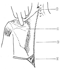
Figure 1 Scapular Depression Surgery
① Severed upper trapezius muscle ② Depressed scapula ③ Skin suture line ④ Excess portion of trapezius muscle


