| disease | Congenital Calcaneovalgus Foot |
Congenital calcaneovalgus foot is a common postural foot deformity characterized by dorsiflexion and eversion of the entire foot. It is more common in females than males, with a ratio of approximately 1:0.6.
bubble_chart Etiology
This condition is more common in first-born infants, possibly related to the primiparous uterus being smaller and having higher tension, along with tighter abdominal muscles. These factors can easily cause mechanical compression on the fetus during the advanced stage of pregnancy, leading to abnormal foot positioning.
bubble_chart Clinical Manifestations
The deformity of dorsiflexion and eversion of the affected foot can be observed immediately after birth. In severe cases, the dorsum of the foot may come into contact with the skin on the anterior aspect of the tibia (Figure 1).
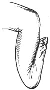
Figure 1 Severe dorsiflexion deformity in congenital calcaneovalgus foot
Due to increased tension in the dorsal and lateral soft tissues, the metatarsus has limited plantarflexion and inversion movements.
X-ray examinations often show no abnormalities, with neither subluxation of the intertarsal joints nor hypoplasia of the primary tarsal ossification centers.
bubble_chart Treatment Measures
The prognosis of this disease is favorable. In mild cases where the affected foot can passively dorsiflex and invert beyond the neutral position, no treatment is required, and it usually resolves spontaneously within 3 to 6 months. For more severe deformities, manual reduction should be performed, involving passive dorsiflexion and inversion exercises to stretch the dorsal and lateral soft tissues of the foot. This should be done 3 to 4 times daily, with 30 repetitions each time. Each stretch should be maintained for about 10 seconds. Generally, persistence for 2 to 3 months can correct the deformity. If manual reduction is unsatisfactory, corrective plaster or a Denis-Brown splint should be applied to fix the affected foot in a dorsiflexed and inverted position, which typically resolves the deformity in approximately 4 to 6 weeks.
bubble_chart DifferentiationThis disease is easy to diagnose, but sometimes it needs to be differentiated from paralytic calcaneovalgus foot caused by neural tube closure defects and congenital vertical talus. The former is characterized by weakened muscle strength in the triceps surae, tibialis posterior, or flexor digitorum longus. X-ray examination may reveal spina bifida below the L3, L4 vertebrae. The latter is caused by dislocation of the talonavicular joint, resulting in a convex pes valgus. Due to the displacement of the talar head toward the metatarsal side, a bony prominence can be palpated on the sole of the foot. A lateral X-ray view may show the talus in a vertical position, with its central axis located posterior to the cuboid and on the metatarsal side.


